
The ability to study cells depends largely on how readily they can be grown and manipulated in the laboratory. Although the process is technically far more difficult than the culture of bacteria or yeasts, a wide variety of animal and plant cells can be grown and manipulated in culture. Such in vitro cell culture systems have enabled scientists to study cell growth and differentiation, as well as to perform genetic manipulations required to understand gene structure and function.
Table of Contents
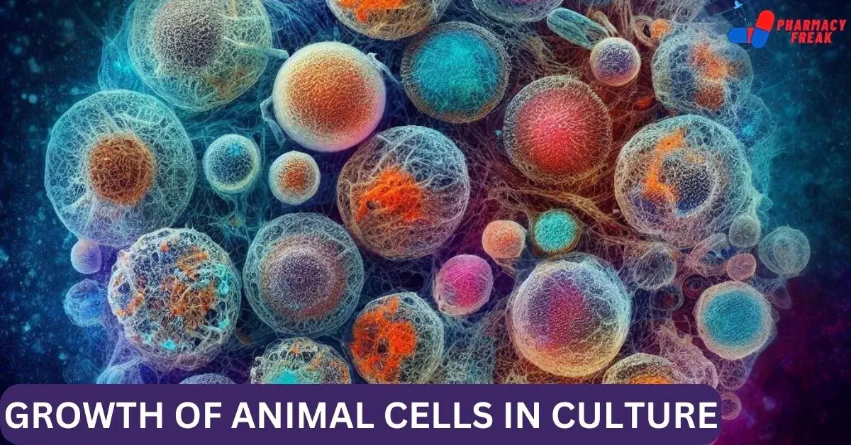
Animal cell cultures start with the dispersion of a piece of tissue into a suspension of its component cells, and then we add it to a culture dish containing nutrient media. Most animal cell types, such as fibroblasts and epithelial cells, attach and grow on the plastic surface of dishes used for cell culture. Because they contain rapidly growing cells, generally we use embryos or tumors as starting material.
Embryo fibroblasts grow particularly well in culture and consequently are one of the most widely studied types of animal cells. Under appropriate conditions, however, some specialized cell types can also be grown in culture, allowing their differentiated properties to be studied in a controlled experimental environment.
Media required for culture of animal cells
The culture media required for the propagation of animal cells are much more complex than the minimal media sufficient to support the growth of bacteria and yeasts. Animal cells grown in culture require nine essential amino acids, namely, histidine, isoleucine, leucine, lysine, methionine, phenylalanine, threonine, tryptophan, and valine. In addition, most cultured cells require cysteine, glutamine, and tyrosine.
Early studies of cell culture utilized media consisting of undefined components, such as plasma, serum, and embryo extracts.
In 1955 Harry Eagle described the first defined media that supported the growth of animal cells. In addition to salts and glucose, the media used for animal cell cultures contain various amino acids and vitamins, which the cells cannot make for themselves.
The growth media for most animal cells in culture also include serum, which serves as a source of polypeptide growth factors that are required to stimulate cell division. Several such growth factors have been identified. They serve as critical regulators of cell growth and differentiation in multicellular organisms, providing signals by which different cells communicate with each other. For example, an important function of skin fibroblasts in the intact animal is to proliferate when needed to repair damage resulting from a cut or wound.
Their division starts with a growth factor released from platelets during blood clotting, thereby stimulating the proliferation of fibroblasts in the neighborhood of the damaged tissue. The identification of individual growth factors has made possible the culture of a variety of cells in serum-free media.
The initial cell cultures established from tissue are primary cultures. The cells in a primary culture usually grow until they cover the culture dish surface. Then we can remove it from the dish and replate it at a lower density to form secondary cultures. We can repeat this process many times, but we cannot grow normal cells in culture indefinitely. For example, we can culture normal human fibroblasts for 50 to 100 population doublings, after which they stop growing and die.
In contrast, cells derived from tumors frequently proliferate indefinitely in culture and hence they are immortal cell lines. In addition, several immortalized rodent cell lines have been isolated from cultures of (normal fibroblasts. Instead of dying as most of their counterparts do, a few cells in these cultures continue proliferating indefinitely, forming cell lines like those derived from tumors. Such permanent cell lines have been particularly useful for many types of experiments. Because they provide a continuous and uniform source of cells that we can manipulate, clone, and indefinitely propagate in the laboratory. Even under optimal conditions, the division time of most actively growing animal cells is approximately 20 hours. Ten times longer than the division time of yeasts.
Consequently, experiments with cultured animal cells are more difficult and take much longer than those with bacteria or yeasts. For example, the growth of a visible colony of animal cells from a single cell takes a week or more, whereas colonies of E. coli or yeast develop from single cells overnight. Nonetheless, genetic manipulations of animal cells in culture have been indispensable to our understanding of cell structure and function.
ANIMAL CELL CULTURE
Cell culture refers to the process by which cells are grown under controlled conditions, generally outside their natural environment i.e. in a controlled artificial environment. These conditions vary for each cell type but generally consist of a suitable vessel with a substrate or medium that supplies the essential nutrients, growth factors, hormones, and gases like carbon dioxide, oxygen; and regulates the physio-chemical environment (pH buffer, osmotic pressure, temperature).
Most cells require a surface or an artificial substrate (adherent or monolayer culture) whereas we can grow others free-floating in a culture medium (suspension culture).
In a cell culture technique, we remove cells from an animal or a plant and grow subsequently in a favorable environment. For animal cell culture, we take the cells from the organ of an experimental animal.
We may remove the cells directly or by mechanical or enzymatic action. We can obtain the cells by previously made cell lines or cell strain. Examples of cells used to culture are fibroblast, lymphocytes, cells from cardiac and skeletal tissues, cells from liver, breast, skin, and kidney, and different types of tumor cells.
TYPES OF ANIMAL CELL CULTURE
Based on the number of cell divisions, we can classify cell culture as primary cell culture and cell lines. Cell lines can undergo finite or infinite cell divisions.
A. Primary cell culture
This is the cell culture obtained straight from the cells of the host tissue. The cells dissociated from the parental tissue are grown on a suitable container and the culture thus obtained is called primary cell culture. Such culture comprises mostly heterogeneous cells and most of the cells divide only for a limited time. However, these cells are much similar to their parents.
Depending on their origin, primary cells grow either as an adherent monolayer or in a suspension.
Adherent cells
These cells are anchorage-dependent and propagate as a monolayer. These cells need to be attached to a solid or semi-solid substrate for proliferation. These adhere to the culture vessel with the use of an extracellular matrix, which is generally derived from tissues of organs that are immobile and imbeembeddeda network of connective tissue. Fibroblasts and epithelial cells are of such types.
When the bottom of the culture vessel is covered with a continuous layer of cells, usually one cell in thickness, these are known as monolayer cultures. The majority of continuous cell lines grow as monolayers. As being single layers, we can transfer such cells directly to a coverslip to examine under a microscope.
Suspension cells
Suspension cells do not attach to the surface of the culture vessels. These cells are also called anchorage-independent or non-adherent cells, which can be grown floating in the culture medium. Hematopoietic stem cells (derived from blood, spleen, and bone marrow) and tumor cells can be grown in suspension. These cells grow much faster do not require the frequent replacement of the medium and can be easily maintained. These are of homogeneous types and enzyme treatment is not required for the dissociation of cells; similarly, these cultures have a short lag period.
Confluent culture and the necessity of sub-culture
After the cells are isolated from the tissue and proliferated under the appropriate conditions, they occupy all of the available substrates i.e. reach confluence. For a few days, it can become too crowded for their container and this can be detrimental to their growth, generally leading to cell death if left for a long time.
The cells thus have to be subculture i.e. a portion of cells is transferred to a new vessel with a fresh growth medium which provides more space and nutrients for the continual growth of both portions of cells. Hence, subculture keeps cells in a healthy and growing state.
A passage number refers specifically to how many times a cell line has been sub-cultured. In contrast with the population doubling level in that, the specific number of cells involved is not relevant. It simply gives a general indication of how old the cells may be for various assays.
B. Secondary cell culture and cell line
When a primary culture is sub-cultured, it is known as secondary culture or cell line, or subclone. The process involves removing the growth media and disassociating the adhered cells (usually enzymatically). Sub-culturing of primary cells into different divisions leads to the generation of cell lines. During the passage, cells with the highest growth capacity predominate, resulting in a degree of genotypic and phenotypic uniformity in the population. However, as they are sub-cultured serially, they become different from the original cell.
Based on the life span of culture, we can categorize the cell lines into two types:
- Finite cell lines
- Continuous cell lines
Finite cell lines
The cell lines, which go through a limited number of cell divisions having a limited life span, we call the minute cell lines. The cells passage several times and then lose their ability to proliferate, which is a genetically determined event known as senescence Cell lines derived from primary cultures of normal cells are finite cell lines.
Continuous cell lines
When a finite cell line undergoes transformation and acquires the ability to divide indefinitely, it becomes a continuous cell line. Such transformation/ mutation can occur spontaneously or can be chemically or virally induced or from the establishment of cell cultures from malignant tissue. Cell cultures prepared in this way can be sub-cultured and grown indefinitely as permanent cell lines and are immortal.
These cells are less adherent, fast-growing, less fastidious in their nutritional requirements, able to grow up to higher cell density, and different in phenotypes from the original tissue. Such cells grow more in suspension. They also tend to grow on top of each other in multilayers on culture-vessel surfaces.
METHODS
Growth Requirements
The culture media used for cell cultures are generally quite complex, and culture condition widely varies for each cell type. However, media generally include amino acids, vitamins, salts (maintain osmotic pressure), glucose, a bicarbonate buffer system (maintains a pH between 7.2 and 7.4), growth factors, hormones, O₂ and CO₂ To obtain the best growth, the addition of a small amount of blood serum is usually necessary, and several antibiotics, like penicillin and streptomycin, are added to prevent bacterial contamination. Temperature varies on the type of host cell.
Most mammalian cells are maintained at 37 degrees Celsius for optimal growth, while cells derived from cold-blooded animals tolerate a wider temperature range (i.e. 15oC to 26°C). Actively growing cells of log phage should be used which divide rapidly during culture.
The process to obtain primary cell culture
Primary cell cultures are prepared from fresh tissues. We remove pieces of tissues from the organ aseptically; which we usually mince with a sharp sterile razor and dissociated by proteolytic enzymes (such as trypsin) that break apart the intercellular cement. Then we wash the obtained cell suspension with a physiological buffer (to remove the proteolytic enzymes used). The cell suspension is spread out on the bottom of a flat surface, such as a bottle or a Petri dish. This thin layer of cells adhering to the glass or plastic dish is overlaid with a suitable culture medium and is incubated at a suitable temperature.
Aseptic techniques
Bacterial infections, like Mycoplasma and fungal infections commonly occur in cell culture creating a problem to identify and eliminate. Thus, all cell culture work is done in a sterile environment with proper aseptic techniques. Work should be done in laminar flow with the constant unidirectional flow of HEPA-filtered air over the work area. All the materials, solutions, and the whole atmosphere should be contamination-free.
Cryopreservation
If a surplus of cells is available from sub-culturing, we should treat it with the appropriate protective agent (e.g., DMSO or glycerol) and store it at temperatures below 130°C. This stores cell stocks and prevent the original cell from being lost due to unexpected equipment failure or biological contaminations. It also prevents finite cells from reaching senescence and minimizes the risks of changes in long-term cultures.
When thawing the cells, we warm the frozen tube of cells quickly in warm water, rinsed with medium and serum, and then added it into culture containers once suspended in the appropriate media.
APPLICATIONS OF CELL CULTURE
1. Vaccines Production
One of the most important uses of cell culture is in the research and production of vaccines. The ability to grow large amounts of virus in cell culture eventually led to the creation of the polio vaccine, and cells are still used today on a large scale to produce vaccines for many other diseases, like rabies, chickenpox, hepatitis B, and measles. In early times, researchers had to use live animals to grow poliovirus, but due to the development of the cell culture technique, they were able to achieve much greater control over virus production on a much larger scale, which eventually developed vaccines and various treatments.
However, we do not use continuous cell lines in virus production for human vaccines as these are derived from malignant tissue or possess malignant characteristics.
2. Cancer Research
Since both normal cells and cancer cells can be grown in culture, the basic differences between them can be closely studied. In addition, it is possible, by the use of chemicals, viruses, and radiation, to convert normal cultured cells into cancer-causing cells. Thus, the mechanisms that cause the change can be studied. Cultured cancer cells also serve as a test system to determine suitable drugs and methods for selectively destroying types of cancer.
3. Virology
One of the earliest and major uses of cell culture is the replication of viruses in cell cultures (in place of animals) for use in vaccine production. Cell cultures are also widely used in the clinical detection and isolation of viruses, as well as basic research into how they grow and infect organisms.
4. Cell-Based Manufacturing
We can use cultured cells to produce many important products; three areas are generating the most interest. The first is the large-scale production of viruses for use in vaccine production. These include vaccines for polio, rabies, chickenpox, hepatitis B, and measles.
The second is the large-scale production of cells that have been genetically engineered to produce proteins that have medicinal or commercial value. These include monoclonal antibodies, insulin, hormones, etc. The third is the use of cells as replacement tissues and organs. Artificial skin for use in treating burns and ulcers is the first commercially available product. However, testing is underway on artificial organs such as the pancreas, liver, and kidney.
A potential supply of replacement cells and tissues may come out of work currently being done with both embryonic and adult stem cells. These are cells that have the potential to differentiate into a variety of different cell types. We hope that learning how to control the development of these cells may offer new treatment approaches for a wide variety of medical conditions.
5. Genetic Counselling
Amniocentesis, a diagnostic technique that enables doctors to remove and culture fetal cells from pregnant women, has given doctors an important tool for the early diagnosis of fetal disorders. We can examine these cells for abnormalities in their chromosomes and genes using karyotyping, chromosome painting, and other molecular techniques.
6. Genetic Engineering
The ability to transfect or reprogram cultured cells with new genetic material (DNA and genes) has provided a major tool to molecular biologists wishing to study the cellular effects of the expression of these genes (new proteins). We can use these techniques to produce these new proteins in large quantities in cultured cells for further study. Insect cells are widely used as miniature cell factories to express substantial quantities of proteins that they manufacture after being infected with genetically engineered baculoviruses.
7. Gene Therapy
The ability to genetically engineer cells has also led to their use for gene therapy. We can remove cells from a patient lacking a functional gene and the missing or then we can replace the damaged gene. We can grow the cells for a while in culture and then we can replace them in the patient. An alternative approach is to place the missing gene into a viral vector and then “infect” the patient with the virus in the hope that the missing gene will then be expressed in the patient’s cells.
8. Screening and Development
Cell-based assays have become increasingly important for the pharmaceutical industry, not just for cytotoxicity testing but also for high throughput screening of compounds that may have potential use as drugs. Originally, these cell culture tests were done in 96 well plates, but increasing use is now being made of 384 and 1536 well plates.


