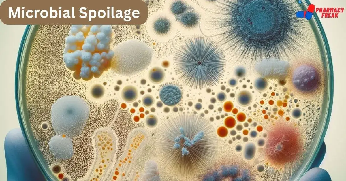
Microorganisms are ubiquitous in the air, food, oil, water, etc, and form an integral part of our environment. Different types of microorganisms mainly contaminate pharmaceutical preparations and spoil them. Such results in major health problems for the users and financial problems for the manufacturer due to the loss of product or expensive litigation with aggrieved users of the medicine.
Table of Contents
In injections, drops, and certain dressings, sterility is essential since any contaminants may cause infections, and the products of contaminants (pyrogens) may cause harmful and even lethal reactions. Oral and topical preparations may cause infections after microbial contamination, the most serious incident being the occurrence of typhoid fever by the ingestion of contaminated thyroid tablets. Excessive microbial contamination may cause more or less extensive deterioration of the product.
Pharmaceutical products may be considered to be microbiologically spoiled if low levels of pathogenic microbes or toxic microbial metabolites are present and detectable physical or chemical changes have occurred in the product.

TYPES OF MICROBIAL SPOILAGE
Types of microbial spoilage are as follow
- Infection induced by contaminated pharmaceutical product
- Physical and ‘chemical deterioration of products
- Observable effects of microbial attack on products
- Ingredients susceptible to microbial attack
- Therapeutic agents
- Surface active agents
- Polymers and humectants
- Fats and oils
- Preservatives and disinfectants
- Sweetening, flavoring, and coloring agents
1. Infection induced by contaminated pharmaceutical product
Pharmaceutical products may be contaminated by pathogenic microorganisms mainly from raw material or at the time of preparations. These contaminated pharmaceuticals may cause serious infections to the patients when they use these drugs or formulations. Cholera in a West African country was traced to an oral liquid drug that had been prepared with contaminated water. Recently, several children died in the UK from septicemia caused by Pseudomonas contamination of parental nutritional fluids during their aseptic preparation. Different dosage forms and contaminated microorganisms that are responsible for diseases are
| DOSAGE FORMS | CONTAMINATING MICROBES | INFECTIONS |
|---|---|---|
| TABLETS/CAPSULES | SALMONELLA SPECIES | SALMONELLA INFECTION |
| EYE DROPS | PSEUDOMONAS AERUGINOSA | EYE INFECTION |
| ANTISEPTIC SOLUTIONS | PSEUDOMONAS SPECIES | SEPTICEMIA |
| OINTMENT AND CREAMS | GRAM-NEGATIVE BACTERIA | DERMATOSES AND BURNS |
| INTRAVENOUS MEDICINES | CANDIDA SPECIES | FATAL SEPTICEMIA |
2. Physical and ‘chemical deterioration of products
Pharmaceutical formulations may be considered specialized micro-environments. Some naturally occurring ingredients are particularly sensitive to attack. Crude vegetable and animal drug extracts often contain wide
assortments of microbial nutrients besides therapeutic agents. The rate of deterioration of ingredients depends upon their chemical structure, the physic-chemical properties of a particular environment, and the level of microbial contamination present.
3. Observable effects of microbial attack on products
Microbial spoilage of different dosage forms may be detected by organoleptic tests. These spoiled products may release very unpleasant smelling and testing metabolites such as ‘sour’ fatty acids, ‘fishy’ amines,” ‘earthy’ or sickly tastes and smells. Formulations may become colored green, pink, brown, black, or yellow by diffusible microbial pigments. Spoiled creams by the microbial attack may become lumpy or gritty. Degradation of surfactant and lowering of pH by lipase attack of triglycerides induces progressive coalescence of lipid droplets and eventually complete separation of the two phases. Surface activity is reduced at a very early stage in the metabolism of most surfactants. Thickening and suspending agents produce a marked reduction in viscosity by depolymerization.
4. Ingredients susceptible to microbial attack
a). Therapeutic agents
Laboratory experiments have demonstrated that many drugs are capable of gross degradation by a wide variety of microorganisms. Potent therapeutic, agents such as analgesics (aspirin, paracetamol), alkaloids (morphine, atropine), barbiturates, steroid esters, etc. can be metabolized and serve as a substance for microbial growth. Aspirin may be converted to salicylic acid and penicillin (by B- lactamase) or chloramphenicol (by chloramphenicol acetylase) to inactive products. Localized transformation of steroids has been observed around fungal colonies growing on the surface of steroid tablets and in steroidal creams. Many microbial transformations are useful for the production of potent steroids.
b). Surface active agents
Alkali-metal and soaps of fatty acids (anionic surfactants) are generally stable due to the slight ph of the formulations, which are easily degraded in sewage. Alkyl and alkylbenzenes sulphonates and sulfate esters are metabolized by – oxidation of their terminal groups followed by sequential B-oxidation of the alkyl chains and fission of the aromatic rings. The cationic surfactants used as antiseptics and preservatives in pharmacies are slowly degraded at high dilution in sewage. Alkypolyoxethlene alcohol emulsifiers (non-ionic surfactants) are readily metabolized by a wide variety of microorganisms. Microbial resistance may be observed by surfactants and are reasonably biodegradable.
c). Polymers and humectants
Thickening and suspending agents used in pharmacy are subjected to microbial depolymerization by extracellular enzymes yielding nutritive .ments and monomers e.g. starch (amylases), pectin (pectinases), dextran (dex. masses), protein (proteases), carboxymethyl cellulose (cellulases) and tragacanth
(oxidases). Polyethylene glycols are readily degraded by sequential oxidation of the hydrocarbon chains but agar (complex polysaccharides) is a relatively inert polymer. Polymers used in plastic packaging are extremely resistant to microbial attack. Glycerol and sorbitol (humectants) in pharmaceuticals readily support microbial growth unless present in high concentration.
d). Fats and oils
Fats and oils (hydrophilic substances are usually attacked extensively when dispersed in aqueous formulations such as oil-in-water droplets contaminate the bulk phase during storage. Lipolytic breakdown of triglycerides emulsions liberates glycerol and fatty acids, then by 3-oxidation of the alkyl chain produces odorous ketones. The microbial metabolism of hydrocarbon oils presents a considerable problem in engineering and fuel technology when water is present as a contaminant.
e). Preservatives and disinfectants
Most organic preservatives and disinfectants are metabolized readily by many bacteria and fungi and may serve as growth substrates at concentrations below their effective use levels. Organomercurial preservatives discharged into rivers from paper mills have been extensively converted to toxic alkyl mercury compounds which could reach humans via an ascending food chain. Degradation of agents at required concentrations in different dosage forms is less commonly reported. Pseudomonas species have metabolized 4-hydroxybenzoate ester preservatives contained in eye drops and caused serious eye infections. Many disinfectants show the growth of microorganisms if these chemicals are diluted.
f). Sweetening, flavoring, and coloring agents
Many sugars and other sweetening agents used in pharmacy are ready substrates for microbial growth. However, some are used in very high concentrations to reduce water activity in aqueous products and inhibit microbial attack. Aqueous stock solutions of flavoring agents such as peppermint water and chloroforms water and coloring agents such as amaranth or tartrazine readily support the growth of bacteria and yeasts.
FACTORS AFFECTING MICROBIAL SPOILAGE
The physical and chemical status of a pharmaceutical formulation influences the type and extent of microbial spoilage considerably. A specific combination of conditions within a product may favor its degradation by a particular group of microorganisms.
Factors affecting microbial spoilage are
- Size of inoculums
- Nutritional factors
- Moisture content
- Temperature
- pH
- Redox potential
- Protective components
1. Size of inoculums
Low levels of contaminants may be present in a product but it would cause low rates of deterioration. Ingredients contaminated by a high level of microorganisms cause appreciable microbial degradation, however, inoculum size ‘alone is not always a reliable indicator of likely spoilage potential. A very low level of Pseudomonas in a weakly preserved solution may suggest a greater risk than tablets containing high numbers of fungal and bacteria spores.
2. Nutritional factors
Gross microbial spoilage of pharmaceutical products generally requires appreciable growth of the contaminating microorganisms within the dosage forms. Most of the organism and inorganic ingredients act as potential carbon tr nitrogen substrates for microbial growth. The complexity of many formulations offers considerable nutritional variety for a wide range of microorganisms. The use of crude vegetable or animal products in a formulation provides an additionally nutritious environment. Demineralized water prepared by ion exchange) contains sufficient”. nutritious to allow the multiplication of water-borne Gram-negative bacteria.
3. Moisture content
Dissolved components of an aqueous formulation may form a complex with water molecules via hydrogen and other bonding thus lowering the proportion of water available to contaminants microorganisms in the product. The high concentration of the solute in the formulation indicates that water activity, (A) is low.
Most microorganisms grow best at high water activity. Hence, formulations can be protected from microbial attack by lowering their water activity with additions of suitable levels of sugars, polyethylene glycols, or sodium chloride or by drying Condensed moisture films can form on the surface of tablets or bulk oils as a result of fluctuations in storage temperatures and the moisture content of the air may be sufficient to initiate fungal growth. Moisture films similarly formed on the surface of viscous syrups permit yeast and fungal growth.
4. Temperature
Spoilage of pharmaceuticals could occur over the range of about -10 to 60°C, although it is much less likely at the extremes. Storage within specific, narrower, temperature ranges will encourage the growth of particular groups of spoilage organisms: Syrups and multidose eye drop preparations are sometimes dispersed with the label ‘store in a cool place, to reduce the risk of in-use contamination before the expiry date. It is recommended that distilled for the preparation of ‘water for injections be stored at 80°C before sterilization, to prevent pyrogen production.
5. pH
Extremes of pH prevent a microbial attack, although the growth of mold is commonly observed in solutions of dilute hydrochloric acid. Antacid mixtures, mouthwashes, and distilled water show growth of Pseudomonas species and other Gram-negative bacteria at neutral pH. Soap emulsion (pH 8.0-9.5) and a mixture of magnesium hydroxide gel discourage microbial attack. Acidic conditions favor fungal and yeast proliferation and inadequately preserved flavored pharmaceutical products at pH 3.5 can support the growth of microbes. Yeast can metabolize acids raising the pH sufficiently for fats and oils.
6. Redox potential
The ability of microbes to grow in an environment is influenced by its oxidation – reduction balance (redox potential), Vacuum packing of foodstuffs, or the inclusion of oxygen absorbers in the package to minimize oxygen levels and reduce
microbial spoilage. Oxygen removed to removal to control spoilage in medicines is not a practical proposition although it is used to control non-biological oxidation. The redox potential even in fairly viscous emulsions may be quite high due to the appreciable solubility of oxygen in most fats and oils.
7. Protective components
Various components of formulations and products may increase the resistance and longevity of contaminant microorganisms. Microbes can show more resistance to heat or desiccation in the presence of some polymers such as starch, acacia, or gelatin. Adsorption of microorganisms onto suspended particles such as kaolin, magnesium trisilicate of aluminum hydroxide gel may increase their longevity. Some such as surfactants, suspending agents and proteins can increase the resistance of micro-organisms to preservatives.
SOURCES AND TYPES OF MICROBIAL CONTAMINATION
The microbiological contamination of pharmaceutical products is influenced by the environment in which they are manufactured and by the materials used in their formulation. Different pharmaceutical formulation may be contaminated by the atmosphere, raw material, water, equipments and human sources and get spoiled.
Sources and types of microbial contamination are
- Atmosphere
- Water
- Raw materials
- Process operators
- Equipments
- Packaging
- Buildings
These sources and types are described as follows:
1.Atmosphere
The number of micro-organisms in the atmosphere depends on the activity in the environment for the growth and multiplication of micro-organisms because it does not contain moisture and nutrients which are required for the growth of microbial cells. Microorganisms are carried into the atmosphere suspended on particles of dust, skin, clothing, droplets of moisture, or sputum following coughing or sneezing. The microbial content of the air may be increased during the handling of contaminated materials during dispensing, blending, and formulations.
The use of starch and some sugars in the dry state may increase the fungi count. Settle plates and different air sampling techniques are commonly used for the isolation and identification of microorganisms present in the atmosphere. Microorganisms commonly isolated from the atmosphere are bacteria and fungi eg. Staphylococcus spp., Streptococcus spp., Clostridium spp., Bacillus spp., Penicillium spp., Aspergillus spp., Mucor spp., Cladosporium spp., etc.
The microbial count of air or atmosphere may be reduced by chemical disinfection, ultraviolet light, filtration and gaseous agents.
2 Water
Water is one of the main constituents of many products and it is also used for washing and cooling process. Different types of microorganisms are present in fresh water and some microbes may contaminate the water from sewage e.g. Pseudomonas spp., Proteus spp., Escherichia coli, Sterptococcus faecalis, Flavobacterium spp., Clostridium spp.,
Chromobacter spp. etc. Softened water is often used to contaminate softened water at the time of its preparation by the base exchange method. Deionized water, distilled water, and water for treatment are commonly used for preparation of different pharmaceuticals. Water may be treated by ultraviolet light, filtration, or stored at elevated temperatures to discourage microbial growth.
3. Raw materials
Raw materials, particularly of natural origin are a potentially rich source of microorganisms. Products from animal sources such as gelatin, desiccated thyroid, pancreas, cochineal, etc. may be contaminated with different pathogens. Some plant-origin materials are used for pharmaceuticals e.g. agar, starch, acacia, tragacanth, etc. these are raw from microbes but incidental microbial contamination may occur. Raw materials mainly contain bacteria and fungi e.g. Bacillus spp., Pseudomonas spp., Lactobacillus spp., Salmonella spp., Cladosporium spp., Fusarium spp., etc. heat treatment, filtration, recrystallization, irradiation or any other sterilization processes are mainly applied for reduction of the microbial load in raw materials.
4. Process operators
Microorganisms may be transferred to pharmaceutical formulations. from process operators. Natural skin flora microorganisms are the main source of contamination. e.g.Staphylococcus aureus, Sarcina spp.. Alcaligens spp., etc. Various dermatophytic fungi such as Epidermophyton spp., Microsporon spp., and Trichophyton spp., may be present. Microorganisms other than the natural skin flora may be transferred from operators as a result of poor personal hygiene.
Wounds showing signs of infection may support Staphylococcus aureus, Staphylococcus pyogenes, Proteus spp., and Pseudomonas aeruginosa. The nasal passages and respiratory tract may contain large numbers of Staphylococcus aureus, Staphylococcus salivarius, Neisseria pharyngitis, Klebsiella pneumonia, etc.
5. Equipments
The equipment should be as simple as possible for the purposes required with a minimum of junctions, valves, and pumps to allow cleaning in place by circulation of detergents or other chemical antimicrobial agents. The types and extent of growth will depend on the source of the contamination, environmental conditions, and nutrients. The efficacy of cleaning in-place systems can be checked by plating out a sample of the final rinsed water nutrient agar or by swab tests.
The in-place cleaning system is mainly used for pipelines, plate and tubular types of heat exchange units, pumps, homogenizers, and all larger equipment. Equipment may be sterilized or disinfected by heat, gaseous agents, or chemicals. Many tanks, small equipment, and reaction vessels are sterilized by steam under pressure (autoclave).
6. Packaging
The microbial flora of packaging materials is dependent upon its composition and storage conditions. Packaging material have a dual role and act both to contain the product and to prevent the entry of microorganisms or moisture. Glass containers, cardboard boxes and closure liners carry different types of micro-organisms if not treated by any methods e.g. Bacillus spp., Penicillium spp., Aspergillus spp., Cladosporium spp., and Micrococcus spp. Dry heat at 170°C is often used for of vials and ampoules which also be sterilized by most heat, chemicals, gaseous agents or radiation techniques.
7. Buildings
Different molds and a few bacteria species are the most common flora of walls and ceilings eg. Aspergillus niger, Aspergillus flavus, Cladosporium spp., Penicillium spp., Bacillus spp., etc. They are mainly found in poorly ventilated buildings. Microorganisms utilize most of their nutrients from the plaster paint. To reduce microbial growth, all walls, floors, and ceilings should be smooth and washable.
In areas where acid or alkaline chemicals or cleaning fluids are applied, a resistant sealing and jointing material must be used. Doors and windows should be fitted flush with walls to prevent entry of dust particles and microorganisms. All windows in manufacturing areas should serve only to permit light entry and not for ventilation. The risk of contamination and spoilage of a pharmaceutical product during formulation, storage, and administration to the patient is generally significant and appreciation of this should be reflected in the formulation and design of the product.
MICROBIAL STANDARDS FOR NON-STERILE PHARMACEUTICALS
Microorganisms-limit standards for non-sterile pharmaceutical dosage forms are given in specific monographs in different pharmacopeias.
The amount of water in the formulation is one of the important factors, mainly considered in establishing the need for microbial standards, Pharmaceutical products have the potential to support microbial growth in the presence of water. Microbial limits for capsules and tablets are generally applied to those of plant, animal, or mineral origins. The susceptibility of particular products to microbial growth and damage is dependent upon the nature of the formulation. Tablets containing natural drugs have a higher microbial count as compared to synthetic drugs.
Most microbial species are Gram-positive, spore-forming, and mold-type. Oral formulation contaminated by micro-organisms mainly causes gastrointestinal disorders eg. Salmonella typhi. External semi-solid preparations applied to the skin, provide the opportunity for Staphylococcus and Pseudomonas and produce localized infections. This topical product should be free from Staphylococcus aureus and Pseudomonas aeruginosa.
ASSESSMENT OF MICROBIAL CONTAMINATION AND SPOILAGE
Assessment of microbial contamination and spoilage are done in following ways.
- Physical and chemical change
- Sterility tests
- Assessment of viable microorganisms in non-sterile products
- Estimation of pyrogens
1. Physical and chemical change
Physical and chemical changes of different pharmaceutical formulations and spoilage. Charge in viscosity, pH, emulsion stability, and loss of surface activity of formulations indicate microbial spoilage. Measurement of oxygen consumption of the product can indicate the degree of oxidative attack and microbial growth.
2. Sterility tests
Materials that are to be tested for sterility mustn’t be subject to contamination from the operator or the environment during the test. For this reason, a sterility test must be conducted by competent and experienced personnel in an adequately clean room with laminar flow cabinet facilities. All injectables and ophthalmic preparations are tested by the sterility test.
3. Assessment of viable microorganisms in non-sterile products
Non-sterile products are tested for viable microorganisms for detection of pathogens and total viable counts. Details of this test are given under “Microbial Limit Tests”.
4. Estimation of pyrogens
A pyrogen is a material that causes a rise in body temperature when injected into a patient. The lipopolysaccharides and lipoproteins that comprise a major part of the cell wall of Gram-negative bacteria are called endotoxins which are the most commonly called pyrogens. Two main procedures are used for the detection of pyrogens(Refer to unit III). The BP pyrogen test requires administration of the injection to laboratory rabbits. Their body temperature is monitored for some time.
The alternative procedure is to use the Limulus Amebocyte Lysate Test (LAL) in which the pyrogen-containing sample causes gel formation in the lysis product of amoebocyte cells of the giant horseshoe crab Limulus polyphemus.
MICROBIAL LIMIT TESTS
The microorganisms quality of non-sterile dosage forms, herbs, processed herbs, and herbal products can be controlled by using two methods.
- Estimation
- Detecting the presence of specific microbial species in pharmaceutical substances
1. Estimation, of the total number of viable aerobic microorganisms in a given sample (total viable count).
2. Detecting the presence of specific microbial species in pharmaceutical substances. The microbial limit test is mainly applied to the raw material of pharmaceutical products of natural or biological origin (e.g. starch, talc, gum, gelatin, etc.) and some finished products (calamine lotion, dried aluminum hydroxide gel, etc).
Detecting species microorganisms from non-sterile dosage forms includes Escherichia coli, Staphylococcus aureus, Pseudomonas aeruginosa, Salmonella, Shigella, Clostridia, and Candida albicans as per Indian Pharmacopeia, 2014.
PRELIMINARY TESTING /ANTIMICROBIAL ACTIVITY OF TEST SAMPLE
The methods given here are invalid if test samples show antimicrobial activity. Hence, first, check the inhibition effect of the test sample by using a viable culture of test microbes. This is done by adding 1 ml of not less than 10 dilutions of 24 hours broth culture of the microorganisms to the first dilution in buffer solution, pH 7.2, fluid soybean casein digest medium, or fluid lactose medium) of the test material and following the test procedure should be modified by:
- Increasing the volume of diluents with the quantity of test material remaining the same
- Incorporating a sufficient quantity of a suitable inactivation agent in the diluents
- Combining the above modifications to permit the growth of the micro-organisms in the media.
If inhibitory substances are present in the sample, 0.5% of soya within and 4% of polysorbate 20 may be added to the culture medium. Alternatively, repeat the test as described above. using fluid casein digest-soya lecithin-polysorbate 20 medium to demonstrate neutralization of preservatives or other antimicrobial agents in the test material Where inhibitory substances are contained in the product and the latter is soluble, the membrane filtration method described under Aerobic Microbial Count may be used.
If all tests fail to inactivate the activity of the test sample it indicates that the article is not likely to be contaminated with the given species of microorganisms.
TOTAL AEROBIC MICROBIAL COUNT
The total count of viable aerobic microorganisms per unit weight or volume of sample is the most commonly used technique in microbiology.
For determination of total aerobic microbial count, it is necessary to pretreat the sample as follows
- Water solution products
- Non-fatty water-insoluble products
- Fatty products
- Fluids or solids in aerosol form
- Transdermal Patches
1. Water solution products
Dissolve 10 gm or 10 ml of the test sample (or as specified in the monograph) in buffered sodium chloride-peptone solution (pH 7.0) or any other suitable medium shown to have no antimicrobial activity under the conditions of the test and adjust the volume to 100 ml with the same medium.
2. Non-fatty water-insoluble products
Suspend a 10 gm or 10 ml test sample (or as specified in the monograph) in a buffered solution chloride-peptone solution (pH 7.0) or any other suitable medium is shown to have no antimicrobial activity under the conditions of the test and adjust the volume to 100 ml with the same medium. A suitable surface active agent (0.1% w/v of ppolysorbate80) may be added to assist in the suspension of poorly wettable substances.
3. Fatty products
Homogenize 10 gm or 10 ml of the sample (or as specified in the monograph) with 5 gm of polysorbate 20 or polysorbate 80. If necessary, heat it to 40°C and mix carefully in the water bath or an oven. Add 85 ml of buffered sodium chloride-peptone (pH 7.0) or any other suitable medium shown to have no antimicrobial activity under the conditions of the test. Maintain the temperature (40°C) for the shortest time formation of an emulsion.
4. Fluids or solids in aerosol form
In sterile conditions, transfer the product into a membrance filter apparatus or to a sterile container for sampling. Use either the total contents or a defined number of material doses, from each of the containers tested. e.
5. Transdermal Patches
Remove the release liners of transdermal patches using sterile forces and place them adhesive side upwards on sterile glass or plastic trays. Cover the adhesive surface with sterile gauze and transfer them to a suitable volume of buffered sodium chloride-peptone solution (pH 7.0) containing an inactivator such as polysorbate 80 or lecithin.
The total aerobic microbial count present in the test substances is determined by the following methods
- Membrance filtration
- Plate count methods
- Pour plate method
- Surface-spread method
- Most probable number (MPN)
1. Membrance filtration
Transfer 10 ml or a quantity of each dilution containing 1 gm of the preparation being examined to each of the two membrance filters (filter, 50 mm diameter, and pore size not greater than 0.45 pm) and filter immediately. If necessary, dilute the pretreated preparation so that the required colony count may be counted. Wash each membrance by filtering through it three or more successive quantities, each of 100 ml of a suitable liquid such as a buffered sodium chloride-peptone solution.
For fatty substances, add to the liquid polysorbate 20 or polysorbate 80. Transfer one of the membrance filters to the surface of the Casein soya bean digest agar plate and the other filter to the surface of the Sabouraud dextrose agar plate with antibiotics. Incubate the plates at 30 to 35°C for 3 to 5 days in the test for bacteria and 20 to 25°C for 5 to 7 days in the test for fungi. Calculate the microorganisms per gm or ml of the test sample.
2. Plate count methods
a). Pour plate method
Mix 1 ml of the pretreated sample and about 15 ml of liquefied Casein soya bean digest agar or Sabouraud dextrose agar plate with antibiotics at not more than 45°C in Petri plates (9 cm diameter). If necessary, dilute the pretreated sample so that a colony count of not more than 300 may be obtained.
Incubate the plates of Casein soya bean digest agar plate at 30 to 35°C for 3 to 5 days and Sabouraud dextrose agar plate at 20 to 25°C for 5 to 7 days for the growth of fungi. Then select plates that have colonies of more than 30 but less than 250 aerobic microorganisms and 20 to 50 colonies of fungi. Calculate the mean count and number of CFU per gm or ml of the product.
b). Surface-spread method
Add 15 ml of liquefied Casein soya bean digest agar er Sabouraud dextrose agar plate with antibiotics in a Petri plate at about 45°C and allow to solidify. Dry the plates in a laminar flow bench or an incubator.
Spread a measured volume of not less than 0.1 ml of the sample prepared as described earlier, over the surface of the medium. Use at least two Petri dishes for each medium and each stain of the test organism. For incubation and calculation of the number of colony-forming units proceed as described in the pour plate method.
3. Most probable number (MPN)
This method is originally known as the multiple-tube or serial dilution method. The procedure and accuracy of the method are less than that of the membrane filtration method or the plate count methods. Hence, this method is carried out when no other method is available.
Prepare a series of at least three subsequent ten-fold dilutions of the product as described in the pre-treatment of the sample, inoculums, and inactivating agents if necessary. From each level of dilution, three aliquots of 1 gm or 1 ml are used to inoculate three tubes with 9 ml of sterile casein soya bean digest broth. Thus, if three levels of dilution are prepared, 9 tubes are inoculated. Incubate all the tubes at 30 to 35°C for 3 days. Record for each level of dilution the number of tubes showing microbial growth.

I am a Registered Pharmacist under the Pharmacy Act, 1948, and the founder of PharmacyFreak.com. I hold a Bachelor of Pharmacy degree from Rungta College of Pharmaceutical Science and Research. With a strong academic foundation and practical knowledge, I am committed to providing accurate, easy-to-understand content to support pharmacy students and professionals. My aim is to make complex pharmaceutical concepts accessible and useful for real-world application.
Mail- Sachin@pharmacyfreak.com

