
Bacteria
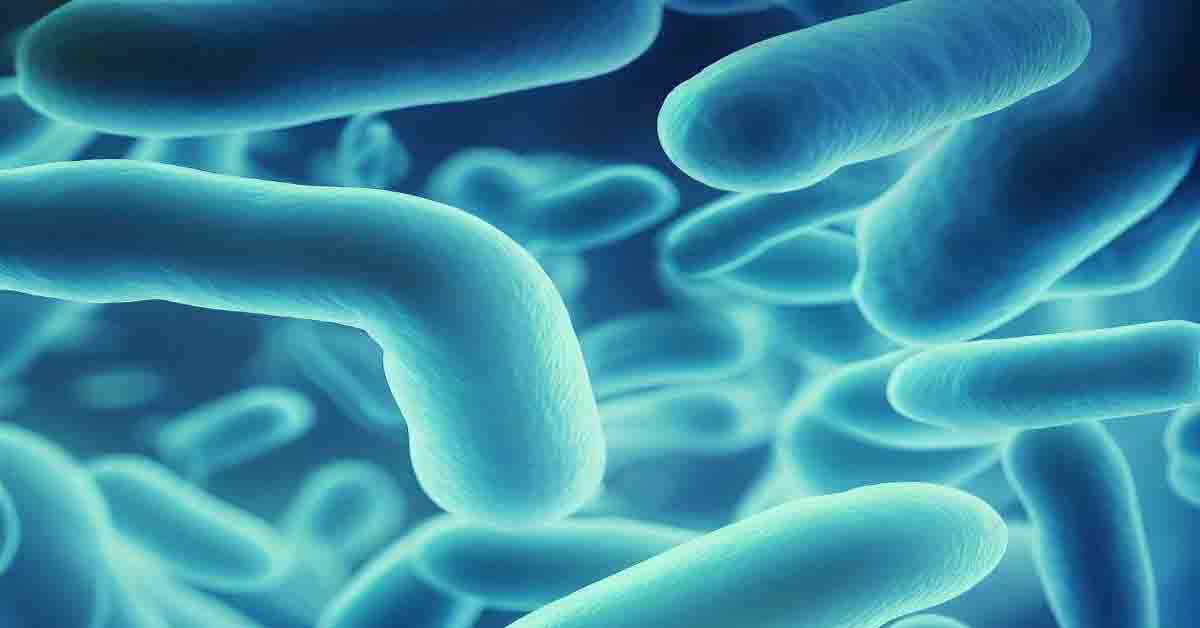
Bacteria (singular: bacterium) form a large group of unicellular prokaryotes that do not contain a nucleus and other membrane-bound organelles. Antony Van Leeuwenhoek first observed them in 1676 using a single-lens microscope of his own design. He called them animalcules” and published his observations in a series of letters to the Royal Society of London. Christian Gottfried Ehrenberg introduced the name bacterium much later in 1838 Typically a few micrometers in length, bacteria have a wide range of shapes, ranging from spheres to rods and spirals.
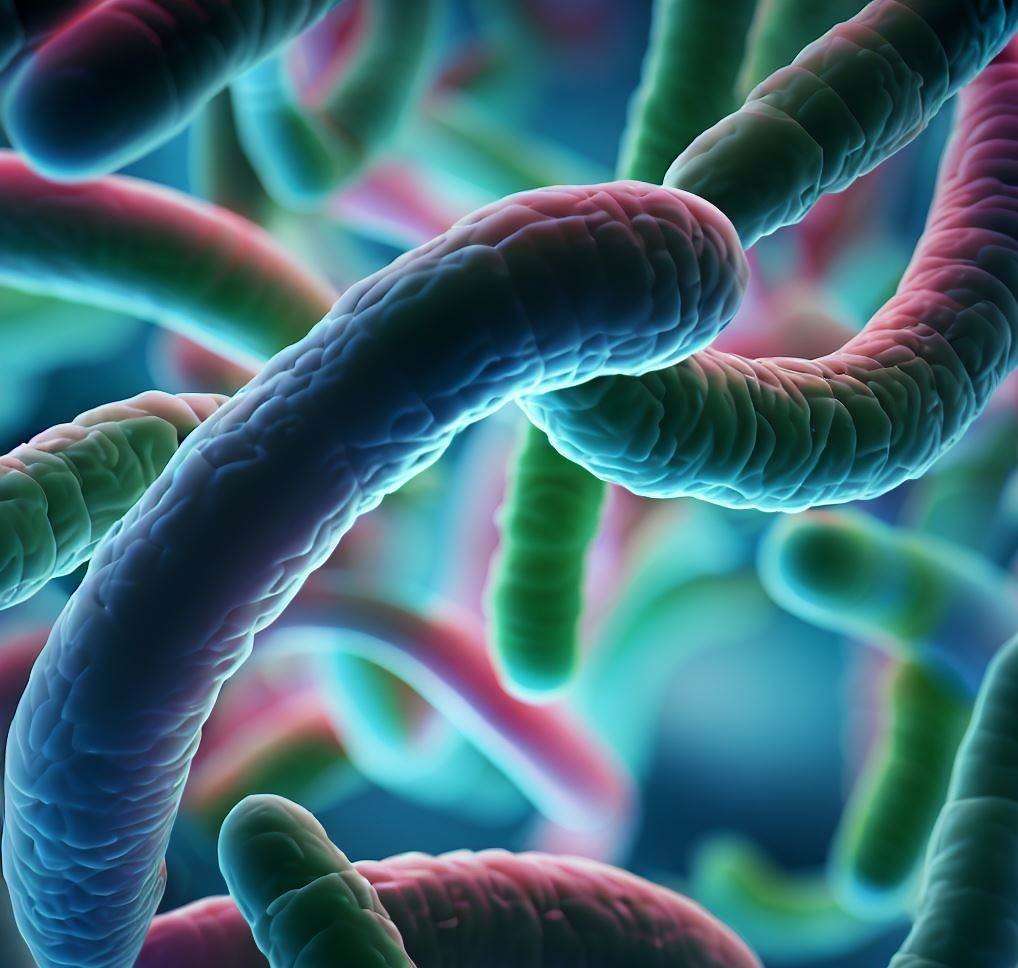
Table of Contents
SIZE OF BACTERIA
Bacterial cells are about one-tenth the sizes of eukaryotic cells and are typically 0.5-5.0 micrometers in length. Medically important bacteria generally measure 0.2-1.5 micrometers in diameter and 3-5 micrometers in length. Among the smallest bacteria are members of the genus Mycoplasma which measures only 0.3 micrometers, as small as the largest viruses. Some bacteria maybe even smaller, but these ultra-micro bacteria are not well studied.
MORPHOLOGICAL CLASSIFICATION OF BACTERIA
Bacteria show great variation in their shapes as discussed below:
Cocci (Greek kokkos – grain or seed)
These are spherical or oval cells. Based on the arrangement of individual organisms they may be;
- Micrococci-cocci– It occurs singly.
- Diplococci-cocci – It occurs in pairs (Diplo means pair).
- Streptococcci-cocci -It occurs in chains (strepto meaning chain).
- Staphylococci-cocci – It occurs in bunches (staphylo meaning bunch).
- Tetrad-cocci – It occur in groups of four (tetra meaning four).
- Sarcinae(octad)-cocci – It occurs in a group of eight(octa meáning eight).
Bacilli (Latin bactilus -stick)
These are rod or stick-shaped cells. Based on the arrangement of an individual organism they may be;
- Microbacilli-bacilli – It occurs singly.
- Diplobacilli- bacilli – It occurs in pairs.
- Streptobacilli-bacilli -It occurs in chains.
- Palisade arrangement – Bacilli lined side by side like matchsticks & at angles to one another e.g. Corynebacterium diphtheriae causing diphtheria.
- Comma-shaped – bacilli that are curved and look like a comma e.g. Vibrio cholera causing cholera.
- Spirillum-bacilli – These are coiled like a cork-screw through 1-5 complete turns e.g., Spirillum minus causing rat-bite fever.
- Coccobacilli– These are the bacteria, which are intermediate to coccus and bacillus e.g., Brucella.
Spirochetes (speir meaning coil; chaise meaning hair)
They are relatively long, slender, flexuous, non-branched microorganisms of spiral shape having several coils e.g., Treponema pallidum causing syphilis.
STRUCTURES EXTERNAL TO CELL WALL
1.FLAGELLA
Flagella (sing: flagellum) are long, hollow, helical filamentous organs of locomotion that arise from the cytoplasmic membrane & pass out through the cell wall. They are 10-20 nm in diameter, 3-20 nm in length & are found on Gram +ve & Gram -ve bacteria. The location of flagella on a cell varies depending upon the bacterial species. They may be polar (at one or both ends of the bacterium) or lateral (along the sides of the bacterium). A flagellum consists of three distinct parts;
- i.Basal body– It constitutes the extreme basal part of the flagellum attached to the plasma membrane.
- ii. Hook– It represents a broader & thicker basal region of the flagellum & passes out through the cell wall.
- iii. Filament– It is the thinner, elongated & terminal part of the flagellum.
The basal body anchored in cytoplasmic membrane comprises a rod & two or more sets of encircling rings-innermost rings (M & S ring) & outermost rings (P & L ring). In Gram-negative bacteria four types of rings, namely M, S, P, and L are present. Through ring M, it attaches to the cell membrane, ring S is located just above cell membrane & through rings P & L; it is attached to peptidoglycan & outer lipopolysaccharide membrane respectively. The outermost rings (P & L ring) are absent in Gram-positive bacteria. The flagellum consists mainly of a protein called flagellin, which belongs to the same chemical groups as myosin (the contractile protein of muscles).
Flagellar types of bacteria
Based on flagellation (pattern of flagellar arrangement) bacteria can be grouped as:
- Monotrichous – It is a bacteria with a single polar flagellum.
- Amphitrichous – It is the bacteria with a single polar flagellum or tuft of flagella at both poles.
- Lophotrichous – It is the bacteria with a tuft of flagella at one pole.
- Peritrichous – It is the bacteria with flagella distributed all around the cell.
Functions: – Flagella help in the motility of bacteria.
2. PILI (FIMBRIAE)
Pili (Sing: pilus) are hollow, non-helical, filamentous appendages that are thinner, shorter & more numerous than flagella. Pili occur in both flagellated & non flagellated bacteria. Each bacterium possesses 100-200 peritrichously-borne (present all around a cell) pili originating from cell membrane. Pili are 1-1.5 um in length & 4-8 nm in diameter & are made up of protein pilin.
Functions:-
- Pili does not function in motility, since they occur in both motile and nonmotile bacteria. There are, however, several functions associated with different types of pili;
- One type of pilus is F-pilus (fertility pilus) or sex pilus as it is involved in bacterial conjugation where it establishes a bridge between a donor & a recipient cell & thus helps in the transfer of genetic material between these bacterial cells.
- Some pili allow the pathogenic bacteria to attach to epithelial cells lining the respiratory, intestinal, or genitourinary tract & thus permit them to establish an infection e.g., Ngonorrhoea adheres specifically to human urethral or cervical epithelium by means of fimbriae & causes a venereal disease-gonorrhea.
3. CAPSULE OR SLIME LAYER
Many bacteria produce substances of high molecular weight at the time of their active growth. These substances collect on the surface of cells & form a gelatinous covering around these cells. When the gelatinous covering does not form a persistent layer but is present more diffusely forming a loose mass or matrix around the bacterial cell, it is a slime layer or a biofilm. This layer is removed by washing the bacterial cell. When the gelatinous covering forms a well-defined persistent (discrete) layer, it is called the capsule. Most bacterial capsules are composed of polysaccharides e.g., capsules of Klebsiella pneumonia, but few capsules are composed of a polypeptide.
e.g. Capsule of Bacillus anthracis.
Functions:
- 1. It protects against temporary drying by binding water molecules.
- 2. It may block the attachment of bacteriophages.
- 3. It may inhibit the engulfment of pathogenic bacteria by phagocytes (WBCs) & thus contributes to virulence (infective ability) of bacteria.
- 4. It may promote attachment of bacteria to smooth surfaces. e.g. Streptococcus mutans, a bacteria which produces dental caries gets attached to the smooth surface of teeth by secreting water-insoluble capsular glucan.
- 5. It may protect the bacteria from antibacterial agents such as lytic enzymes found in nature.
- 6. The capsular material (e.g. dextrans) which may be overproduced when bacteria are fed sugars, becomes reserves of carbohydrates for subsequent metabolism overproduced
4. CELL WALL
It is a rigid structure surrounding the plasma membrane & is present in all Prokaryotes except for Mycoplasma & Methanoplasma.
Structure & chemical composition:-
The main constituent or backbone of the bacterial cell wall is peptidoglycan (also known as murein, muramic acid, or mucopeptide) which is an insoluble, porous, cross-linked polymer of enormous strength & rigidity. Peptidoglycan is found only in prokaryotes, where it occurs in the form of a bag-shaped macromolecule’ surrounding the cytoplasmic membrane. Peptidoglycan consists of two parts:
- A peptide portion – Composed of four amino acids (L-alanine, D-glutamine, either L lysine or diaminopimelic acid & D-alanine) connected by peptide-linkages & hence is called tetrapeptide chain. The two adjacent tetrapeptide chains are interlinked by a cross-linkage (Peptide Bridge).
- Glycan or sugar portion – Made up of alternating units of N-acetyl glucosamine (NAG) & N-acetyl muramic acid (NAM) bonded with each other by ß-1, 4-linkages. Peptidoglycan is present in Gram +ve & Gram -ve bacterial cell walls. However, the cell wall of Gram +ve bacteria differs markedly from that of Gram -ve bacteria in the following perspectives:
Cell wall of Gram +ve bacteria:-
The cell wall of Gram +ve bacteria appears as a thick homogenous layer & mainly consists of peptidoglycan (50% or more of the dry weight of the cell wall), proteins, polysaccharides & teichoic acid. Teichoic acids are acidic polysaccharides that lie on the outer surface of the peptidoglycan and affect the passage of ions thereby helping to maintain the cell wall at a relatively low pH so that autolysins do not degrade the cell wall. They also act as receptor sites for bacteriophages.
Cell Wall of Gram’ -ve Bacteria
The cell wall of Gram’ -ve bacteria is more complex than those of Gram + ve bacteria are. The unique difference is the presence of an outer membrane that surrounds a thin underlying layer of peptidoglycan. Because of this outer membrane, the cell wall of Gram -ve bacteria is rich in lipids (10-25% of the dry weight of the cell wall). The outer membrane is attached to underlying peptidoglycan by means of Braun’s lipoprotein. The membrane is bilayered consists mainly of phospholipids, proteins & lipopolysaccharides (LPS). LPS has toxic properties & is also known as endotoxin, which is released only after disintegration or lysis of the cell.
The outer membrane serves as an impermeable barrier to prevent the escape of important enzymes (involved in cell wall growth) from the space between the cell membrane & the outer membrane (periplasmic space). The membrane also prevents the entry of various external chemicals & enzymes that could damage the cell. Although impermeable to large molecules like protein, the outer molecule allows the smaller molecule (amino acids, peptides, nucleotides, etc.) to pass across using channels in special proteins called porins.
Functions:-
- 1. The cell wall is a rigid structure, that gives shape to the cell.
- 2. Cell wall prevents the cell from expanding & bursting when osmotic pressures are exerted on it.
STRUCTURES INTERNAL TO CELL WALL
1.CYTOPLASMIC MEMBRANE/PLASMA MEMBRANE:
Immediately beneath the cell wall is a plasma membrane, approximately 7.5 nm thick & is composed of phospholipids (about 20-30%) & proteins (about 60-70%). The ultrastructure of the cell membrane was explained by Fluid Mosaic Model proposed by Singer & Nicolson (1974). The phospholipids form a bilayer in which most of the proteins are firmly held as integral or intrinsic proteins, which can be removed only by the destruction of the membrane. Other proteins called peripheral or extrinsic proteins are loosely attached & can be removed by mild treatments such as osmotic shock.
Functions:
- The plasma membrane acts as a differentially permeable barrier, regulating the flow of materials in & out of the cell. Specific proteins (transport proteins) in the membrane allow/facilitate the passage of small molecules (nutrients & waste products) across the membrane.
- It contains various enzymes involved in respiratory metabolism & in the synthesis of cell walls, septum formation, membrane synthesis & DNA replication.
- It is the site of the generation of proton motive force (the force that drives ATP synthesis from ADP, certain nutrient transport systems & flagellar motility).
- It contains specific attachment sites for chromosomes & for plasmid that plays a vital role at the time of cell division.
2.CYTOPLASM
It is a homogenous aqueous solution bounded by cell membrane & is divided into three distinct areas.
- i. Cytoplasmic area – It is Granular in appearance & rich in ribosomes (70S-50S & the 30S) on which proteins are synthesized.
- ii. Chromatin area – It is Rich in DNA.
- iii. Fluid portion – It Consists of dissolved substances (cell solutes, metabolites, inorganic ions).
Unlike higher eukaryotic cells (animals & plants), the bacterial cytoplasm lacks Endoplasmic reticulum, Golgi apparatus, Mitochondria & a true membrane-bound nucleus.
3.INCLUSIONS
Often contained in the cytoplasm of prokaryotic cells is one or another type of inclusion granule. Inclusions are distinct granules that may occupy a substantial part of the cytoplasm. Inclusion granules are usually reserved materials of some sort. For example, carbon and energy reserves may be stored as glycogen (a polymer of glucose) or as polybetahydroxy butyric acid (a type of fat) granules.
Polyphosphate (volutin or metachromatic granules) inclusions are reserves of phosphate and possibly energy; elemental sulfur (sulfur globules) are stored by some phototrophic and some lithotrophic prokaryotes as reserves of energy or electrons. Some inclusion bodies are membranous vesicles into the cytoplasm which contain photosynthetic pigments or enzymes. Some bacteria, which live in aquatic habitats, form gas vacuoles that provide buoyancy to them in aquatic environments.
4.MESOSOMES
Many bacteria especially Gram-positive bacteria possess characteristic membrane invaginations or infoldings in the form of a system of convoluted tubules & vesicles known as mesosomes.
On the basis of their location in a cell they may be:-
- Central:Central mesosomes penetrate deeply into the cytoplasm, are located near the middle of the cell & appear to be attached to the nuclear material of the cell. They are thought to be involved in DNA replication & septa formation at the time of cell division.
- Peripheral: Peripheral mesosomes do not penetrate into the cytoplasm & are located near the periphery. They are thought to be involved in the export of exocellular enzymes like penicillinase.
5. NUCLEAR MATERIAL
Bacterial cells contain neither a distinct membrane-bound nucleus nor a mitotic apparatus. However, they do certain an area near the center of the cell that is regarded as a nuclear structure. Because it is not a discrete nucleus, many names like nucleoid, the chromatin body, the nuclear equivalent & even the bacterial chromosome designated to it. The nucleoid is typically one large circular molecule of DNA, more or less free in the cytoplasm although coiled & supercoiled anchored by proteins. Sometimes a smaller extrachromosomal piece of DNA is present in addition to a nucleoid called a plasmid. The total DNA content of a prokaryote is referred to as a cell genome. The DNA is visible under a light microscope by feulgen staining which is specific for DNA.
6. ENDOSPORES
Certain bacterial smant structures called spores. As these spores are formed within the parent bacterial cell, they are called endospores. They are thickly walled, highly refractile bodies that are usually produced by cells growing in rich culture media but which are approaching the end of active growth. Endospores are produced (one per cell) by certain species of Bacillus (B.anthracis, B.subtilis), Clostridium (Cl. tetani, Cl.welchii & Cl. botulinum), Sporosarcina, Thermoactinomyces & few other genera.
Morphology of spore
The endospore consists of following parts:-
- The innermost layer of the spore wall is the germ cell membrane, inner membrane, or core membrane that surrounds the core.
- The spore wall synthesizes a thick covering layer, the cortex which contains an unusual type of peptidoglycan sensitive to lysozyme.
- Outside the cortex is a densely stained coat (called spore coat) which may be differentiated into an outer coat layer & inner coat layer. The spore coat is made up of keratin-like protein.
- Spores of some species (e.g., B. thuringensis) have an additional loose outer covering called exosporium.
Formation of Spore (Sporulation):
Spore formation is initiated by the appearance of a clear area în a portion of protoplasm near one end of the bacterial cell, which incorporates part of the nuclear material equivalent to one genome of the cell. This clear area with nuclear material becomes gradually more opaque with condensation of nuclear chromatin forming the forespore. The cell membrane grows inwards & undergoes enfolding forming a double-layered membrane structure (spore wall) around the core (forespore). The innermost layer of the spore wall forms the spore membrane. The spore wall then synthesizes other layers – spore cortex, spore coats & exosporium.
Shape & position of spores:
Spores may be spherical or oval & may be central, sub terminal or terminal in position.
Resistance
The endospores are resistant to ordinary boiling, heating & disinfectants. They can withstand boiling up to 3 hours, dry heat at 150 °C for 1 hour. However, they can be destroyed by autoclaving at 121 °C for 15-20 minutes.
All endospores contain large amount of dipicolinic acid (DPA), a unique compound that accounts for 10-15% dry weight of spore. It occurs in combination with large amounts of calcium & this Ca-DPA complex is thought to play a major role in the heat resistance of endospores. a
Germination of spore
The conversion of the spore into a vegetative cell under favorable environmental conditions is known as germination. It may occur in less than 2 hours under optimal conditions.
It consists of following three stages:
- Activation: The activation of the spore is brought about by one or another agent such as heat (60°C for 1 hour), low pH (acidic), abrasion, etc. that damages the spore coat.
- Initiation: Once activated, the process of initiation begins marked by binding of effector substances from a rich medium to the spore coat. This binding activates autolysins (self-produced lysing enzymes) that destroy the peptidoglycan of the cortex, allowing the uptake of water & release of calcium dipicolinate. The number of hydrolytic enzymes produced degrades the various constituents of the spore. self-produced
- Outgrowth: With the disintegration of the cortex & swelling of spore, a single germ cell emerges after breaking open the spore coat. The new vegetative cell consists of the spore protoplast with a surrounding wall. This is followed by a period of active biosynthesis producing an outgrowth that ultimately gets transformed into a new vegetative cell.
Difference between GRAM POSITIVE & GRAM NEGATIVE BACTERIA
| GRAM- POSITIVE BACTERIA | GRAM-NEGATIVE BACTERIA |
| 1. Retains crystal violet dye and appear dark violet or purple | 1. Do not retairi crystal violet, take counter stain (safranin) and appear red |
| 2. Peptidoglycan layer is thick (multi layered) | 2. Peptidoglycan layer is thin (single layered). |
| 3. Teichoic acids are present in many | 3. Absent |
| 4. Periplasmic space is absent | 4. Present |
| 5. Outer membrane is absent | 5. Present |
| 6. Lipopolysaccharide (LPS) content is virtually none | 6. High |
| 7. Lipid and lipoprotein content is low | 7. High (due to presence of outer membrane). |
| 8. Possess 2 rings in basal body (M & S) | 8. Possess 4 rings in basal body (M, S, P& L). |
| 9. Primarily produce exotoxins | 9. Primarily produce endotoxins |
| 10. Inhibited by basic dyes | 10. Usually not inhibited |
| 11. Resistance to physical disruption is high | 11. Low |
| 12 Resistance to sodium azide is high | 12. Low |
| 13. Resistance to drying is high | 13. Low |
| 14. Nutritional requirement is relatively complex | 14. Relatively simple |
| 15. Mesosomes present | 15. Rare or absent |
Differentiate between FLAGELLA & FIMBRIAE
| FLAGELLA | FIMBRIAE |
| 1. They are thicker & larger in size | 1. Thinner & smaller in size |
| 2.They are helical and never straight | 2. Non-helical & always straight |
| 3. They arise from cytoplasmic membrane but not attached to cell wall | 3. Attached to cell wall |
| 4. They are present only in motile species | 4. Present in both motile & non-motile species |
| 5. They are organs of movement( motility) | 5. Organs of adhesion and conjugation |
Differentiate between CELL WALL OF GRAM POSITIVE & CRAM NEGATIVE BACTERIA
| Gram +ve cell wall | Gram-ve cell wall |
| 1.Thick & homogenous | 1.Thin & usually tri-layered. |
| 2. Peptidoglycan comprises up to 90% of cell wall. | 2 Peptidoglycan comprises only 10% or less of cell wall |
| 3.Cell wall rigid | 3. Less rigid |
| 4.Besides peptidoglycan, there are teichoic acids other polysaccharides & proteins in cell wall. | 4.Besides peptidoglycan, there are phospholipids, proteins & lipopolysaccharides in cell wall. |
| 5.Teichoic acids are main surface antigens | 5. Lipopolysaccharides are main surface antigens. |
| 6.More sensitive to wall attacking antibiotics | Less sensitive to wall attacking antibiotics |
| 7. No toxic property of cell wall | 7.Cell wall contains LPS which are endotoxins. |
This is all about bacteria in microbiology.
.


