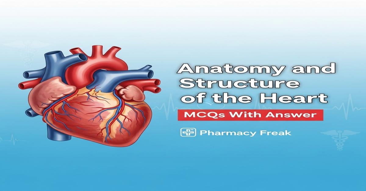
Understanding the anatomy and structure of the heart is essential for B. Pharm students because cardiac architecture underpins pharmacodynamics, drug delivery, and cardiovascular therapeutics. This concise review covers cardiac chambers, valves, myocardial layers, conduction system, coronary circulation, heart surfaces and the fibrous skeleton, integrating gross anatomy with histology and clinical correlations. Emphasis on valve mechanics, nodal and conduction pathways, coronary arterial supply, and fetal circulatory remnants equips pharmacy students to appreciate drug targets, adverse effects and hemodynamic consequences. Clear familiarity with these structures aids interpretation of ECG basics, drug actions on cardiac tissue, and safe medication use in cardiac disease. Now let’s test your knowledge with 30 MCQs on this topic.
Q1. Which cardiac chamber has the thickest myocardial wall?
- Right atrium
- Right ventricle
- Left atrium
- Left ventricle
Correct Answer: Left ventricle
Q2. Which valve separates the left atrium from the left ventricle?
- Tricuspid valve
- Pulmonary valve
- Mitral (bicuspid) valve
- Aortic valve
Correct Answer: Mitral (bicuspid) valve
Q3. The “widow-maker” artery commonly refers to which coronary vessel?
- Right coronary artery (RCA)
- Left circumflex artery (LCX)
- Left anterior descending artery (LAD)
- Posterior descending artery (PDA)
Correct Answer: Left anterior descending artery (LAD)
Q4. Which structures anchor the atrioventricular valve leaflets to the ventricular wall?
- Paget fibers
- Chordae tendineae
- Trabeculae carneae
- Endocardial folds
Correct Answer: Chordae tendineae
Q5. Where is the sinoatrial (SA) node located?
- Interventricular septum near apex
- Roof of left atrium near pulmonary veins
- Anterior wall of right ventricle
- Superolateral wall of right atrium near the SVC opening
Correct Answer: Superolateral wall of right atrium near the SVC opening
Q6. The atrioventricular (AV) node is typically located in which area?
- Anterior interventricular sulcus
- Lower interatrial septum near the coronary sinus
- Posterior left ventricular wall
- At the apex of the heart
Correct Answer: Lower interatrial septum near the coronary sinus
Q7. The bundle of His divides into right and left bundle branches located in the:
- Interatrial septum
- Epicardium over the left ventricle
- Subendocardial layer of the atria
- Interventricular septum
Correct Answer: Interventricular septum
Q8. The fibrous skeleton of the heart primarily consists of:
- Smooth muscle bundles surrounding the coronary arteries
- Dense irregular connective tissue around the valves
- Endothelial cell layers lining the chambers
- Adipose tissue in the atrioventricular groove
Correct Answer: Dense irregular connective tissue around the valves
Q9. The visceral layer of the serous pericardium is also known as the:
- Endocardium
- Fibrous pericardium
- Epicardium
- Myocardium
Correct Answer: Epicardium
Q10. Which is the correct order of heart wall layers from outermost to innermost?
- Endocardium → Myocardium → Epicardium
- Epicardium → Myocardium → Endocardium
- Myocardium → Epicardium → Endocardium
- Pericardium → Endocardium → Myocardium
Correct Answer: Epicardium → Myocardium → Endocardium
Q11. Most venous blood from the myocardium drains into the right atrium via the:
- Superior vena cava
- Coronary sinus
- Great saphenous vein
- Pulmonary veins
Correct Answer: Coronary sinus
Q12. Which valve typically has three cusps?
- Mitral valve
- Tricuspid valve
- Aortic valve
- Pulmonary valve
Correct Answer: Tricuspid valve
Q13. How many papillary muscles are usually present in the left ventricle?
- One
- Two
- Three
- Four
Correct Answer: Two
Q14. Which septum separates the right and left atria?
- Interventricular septum
- Interatrial septum
- Atrioventricular septum
- Membranous septum of the ventricles
Correct Answer: Interatrial septum
Q15. In fetal circulation, the foramen ovale connects which two chambers?
- Right ventricle and left ventricle
- Left atrium and left ventricle
- Right atrium and left atrium
- Right atrium and right ventricle
Correct Answer: Right atrium and left atrium
Q16. The ductus arteriosus connects which two vessels in the fetus?
- Aorta and superior vena cava
- Pulmonary trunk and aorta (descending)
- Pulmonary veins and left atrium
- Right ventricle and pulmonary veins
Correct Answer: Pulmonary trunk and aorta (descending)
Q17. The first heart sound (S1) is mainly caused by closure of the:
- Closure of the atrioventricular (mitral and tricuspid) valves
- Opening of the mitral valve
- Closure of the coronary sinus valve
Correct Answer: Closure of the atrioventricular (mitral and tricuspid) valves
Q18. Parasympathetic innervation to the heart is primarily provided by which nerve?
- Phrenic nerve
- Vagus nerve (CN X)
- Sympathetic trunk
- Glossopharyngeal nerve (CN IX)
Correct Answer: Vagus nerve (CN X)
Q19. Which coronary artery most commonly supplies the sinoatrial (SA) node?
- Left anterior descending (LAD)
- Left circumflex (LCX)
- Right coronary artery (RCA)
- Great cardiac vein
Correct Answer: Right coronary artery (RCA)
Q20. One major electrical role of the cardiac fibrous skeleton is to:
- Conduct impulses faster than Purkinje fibers
- Electrically insulate the atria from the ventricles
- Generate pacemaker potentials
- Store calcium for contraction
Correct Answer: Electrically insulate the atria from the ventricles
Q21. Which great vessel arises from the right ventricle?
- Aorta
- Pulmonary trunk
- Superior vena cava
- Inferior vena cava
Correct Answer: Pulmonary trunk
Q22. The cardiac apex is formed primarily by which chamber?
- Right atrium
- Right ventricle
- Left atrium
- Left ventricle
Correct Answer: Left ventricle
Q23. Cardiac myocytes are electrically coupled by specialized junctions called:
- Desmosomes only
- Tight junctions
- Intercalated discs (including gap junctions)
- Haversian canals
Correct Answer: Intercalated discs (including gap junctions)
Q24. The aortic valve is best auscultated at which surface landmark?
- Left 2nd intercostal space at midclavicular line
- Right 2nd intercostal space at the sternal border
- Left 5th intercostal space at midaxillary line
- Right 4th intercostal space at midclavicular line
Correct Answer: Right 2nd intercostal space at the sternal border
Q25. Ventricular septal defects (VSDs) most commonly occur in which part of the septum?
- Muscular interventricular septum
- Membranous part of the interventricular septum
- Interatrial septum
- Atrioventricular septum
Correct Answer: Membranous part of the interventricular septum
Q26. The coronary ostia (openings) are located in the:
- Left ventricular apex
- Aortic sinuses (sinuses of Valsalva)
- Pulmonary trunk just distal to semilunar valves
- Superior vena cava
Correct Answer: Aortic sinuses (sinuses of Valsalva)
Q27. Purkinje fibers are primarily located in which layer of the heart?
- Epicardium
- Mid-myocardium
- Subendocardial layer
- Pericardial cavity
Correct Answer: Subendocardial layer
Q28. Prominent muscular ridges found on the inner ventricular surfaces are called:
- Pectinate muscles
- Trabeculae carneae
- Chordae tendineae
- Coronary sulci
Correct Answer: Trabeculae carneae
Q29. Which heart valve is most commonly affected by calcific stenosis in older adults?
- Tricuspid valve
- Pulmonary valve
- Aortic valve
- Mitral valve (bicuspid)
Correct Answer: Aortic valve
Q30. Which chamber receives oxygenated blood from the pulmonary veins?
- Right atrium
- Right ventricle
- Left atrium
- Left ventricle
Correct Answer: Left atrium

I am a Registered Pharmacist under the Pharmacy Act, 1948, and the founder of PharmacyFreak.com. I hold a Bachelor of Pharmacy degree from Rungta College of Pharmaceutical Science and Research. With a strong academic foundation and practical knowledge, I am committed to providing accurate, easy-to-understand content to support pharmacy students and professionals. My aim is to make complex pharmaceutical concepts accessible and useful for real-world application.
Mail- Sachin@pharmacyfreak.com
