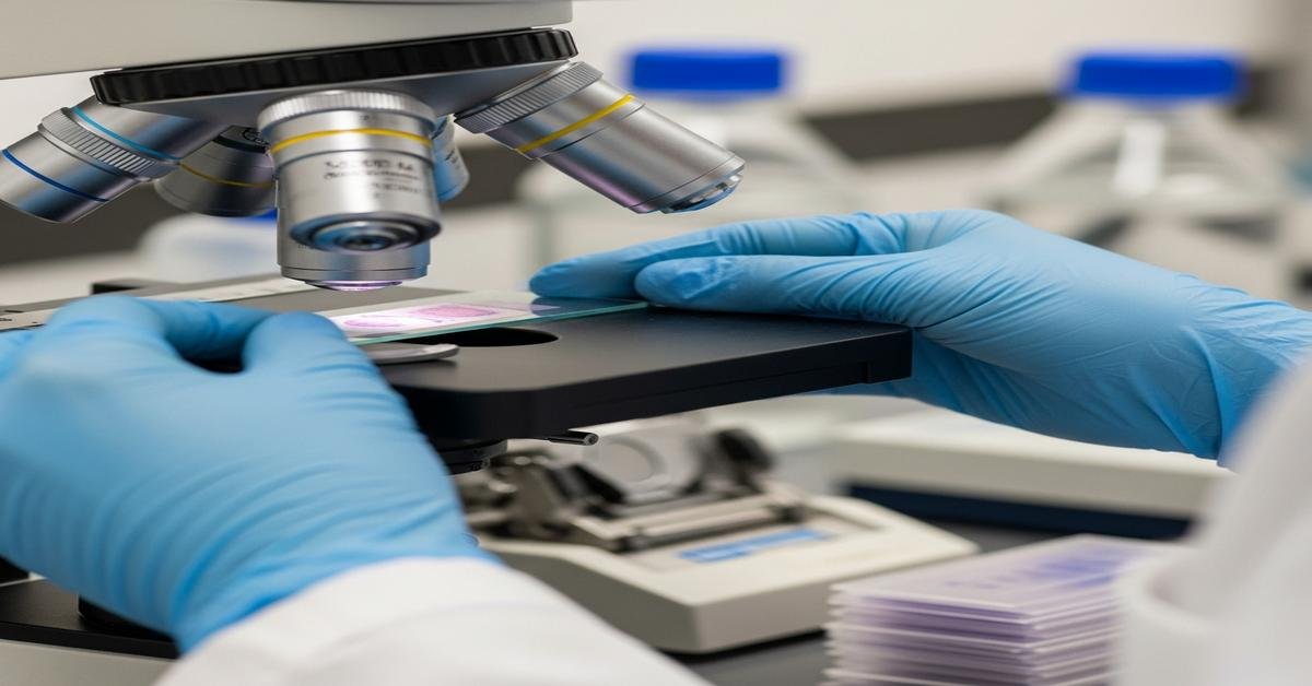
Histotechnology turns tissue into answers. As an HT (ASCP) histotechnician, you fix, process, cut, and stain tissue so pathologists can diagnose disease. The work is exact, fast, and unforgiving. Small mistakes become big artifacts under the microscope. This article explains the science you must master and a clear plan to pass the ASCP Histotechnician board exam on your first try.
What a Histotechnician Really Does
Histotechs keep the diagnostic engine running. You stabilize tissue, move water out and paraffin in, embed for orientation, cut thin sections, and stain for contrast or specific targets. Why this matters: every downstream decision—cancer staging, margins, therapy—depends on your slides.
- Fixation: Preserves structure by stopping autolysis and putrefaction. Done right, nuclei are crisp and cytoplasm is clean. Done poorly, tissue smears, washes out, or overhardens.
- Processing: Dehydrates, clears, and infiltrates. This prevents collapsed tissue and permits thin, intact ribbons.
- Embedding & orientation: Places epithelium on edge, skin on a flat plane, needle cores parallel. The “why”: correct orientation shows the diagnostic surface.
- Microtomy: Produces 3–5 µm sections with intact morphology. Thickness and angle matter because the blade shears, not plows.
- Staining: Adds chemical contrast. H&E reveals architecture; special stains and IHC highlight organisms, fibers, metals, mucins, antigens.
The HT (ASCP) Exam at a Glance
The exam is computer-based, typically about 100 multiple-choice questions in roughly 2.5 hours. ASCP uses a scaled score (0–999). A score of 400 is passing. Content emphasizes fixation, processing, embedding/microtomy, routine and special stains, IHC fundamentals, lab operations, and safety. Staining and microtomy carry the most weight because they dominate daily practice and are the major sources of error.
Eligibility routes usually include one of the following: completion of a NAACLS-accredited HT program; or college coursework in biology and chemistry plus clinical histology experience; or specified on-the-job training. Requirements can change, so verify the current ASCP content outline and route details before you apply. Knowing your route matters because it shapes what experience gaps you need to study harder.
Core Science You Must Master
Focus on concepts that explain outcomes at the bench. When you know the “why,” troubleshooting becomes automatic.
- Fixation
- 10% neutral buffered formalin (NBF): About 4% formaldehyde in phosphate buffer. Crosslinks proteins (methylene bridges) to preserve structure. Buffering prevents acid formaldehyde hematin pigment. Adequate fixation (6–24 hours for most tissues; thinner fixes faster) reduces autolysis and improves nuclear detail.
- Coagulant fixatives: Alcohols (ETOH), acetone, mercuric chloride (historical) precipitate proteins. They penetrate fast and preserve nucleic acids well but can cause shrinkage.
- Glyoxal, Bouin’s (picric acid), zinc formalin: Modify chemistry for particular goals (crisper H&E, better glycogen, reduced mercury hazards). Bouin’s improves soft tissues but can over-picrate and requires picric removal with lithium carbonate or 50–70% alcohol.
- Artifacts & prevention:
- Formalin pigment: Acidic formalin + blood-rich tissue. Prevent with buffered formalin. Remove with saturated alcoholic picric acid.
- Autolysis: Delay to fixation or large specimens. Solve by slicing to 3–5 mm and fixing promptly at a 10–20:1 fixative:tissue volume.
- Overfixation: Excessively hard blocks; weaker antigenicity for IHC. Use appropriate times; for IHC, validate fixation windows.
- Processing
- Dehydration: Graded alcohols (70–95–100%). Why graded? To avoid rapid extraction that collapses tissue.
- Clearing: Xylene or substitutes. These are miscible with alcohol and paraffin. Incomplete clearing leads to mushy blocks and poor ribboning.
- Infiltration: Paraffin at 56–60°C. Overheating causes brittleness and stain loss; under-infiltration causes holes and chatter.
- Decalcification: Strong acids (HCl, nitric) are fast but damage nuclei; chelators (EDTA) are slow but gentler. Always verify endpoint (x-ray, chemical test, or tactile bend test) to prevent under/over-decalcification.
- Embedding
- Orientation: Skin epidermis down; GI mucosa on edge; prostate cores straight; biopsies flat. Orientation determines what the pathologist sees first.
- Temperature: Keep mold, tissue, and paraffin warm enough to prevent interface lines. Quick, even cooling (cold plate) reduces crystallization.
- Microtomy & Cryotomy
- Section thickness: Usually 3–5 µm for H&E; thinner for kidney or bone marrow biopsies. Thinner sections improve nuclear detail but risk chatter on hard tissue.
- Angles: Clearance angle ~3–8°. Too high = chatter; too low = compression. The “why”: the blade must slice, not scrape.
- Water bath: 40–45°C (about 10–15°C below paraffin melting point). Too hot expands sections and weakens staining.
- Frozen sections: Cryostat around –20 to –25°C for most tissue; fattier tissue needs colder. Dull blades and warm blocks cause compression and curling.
- Hematoxylin & Eosin (H&E)
- Hematoxylin: Basic dye + mordant (aluminum or iron) binds nucleic acids. Overstain then differentiate for crisp nuclei. Bluing (alkaline water or ammonia) converts to the blue form. If nuclei are red, bluing was inadequate.
- Eosin: Acid dye stains proteins and RBCs pink to red. pH controls intensity; higher pH weakens eosin. Alcohol carryover dilutes eosin; water carryover scatters it.
- Common errors: Pale nuclei (underfixation or exhausted hematoxylin), muddy background (inadequate dehydration/clearing), uneven stain (dirty reagents or poor agitation).
- Special Stains
- PAS: Oxidizes glycogen and mucins to aldehydes; magenta with Schiff. Diastase digestion confirms glycogen. Why it matters: differentiates fungal walls and basement membranes too.
- GMS (Grocott’s methenamine silver): Fungal walls black on green background. Requires oxidation (chromic acid), silver impregnation, and careful toning. Over-oxidation weakens signal.
- AFB (Ziehl–Neelsen/Kinyoun): Acid-fast bacilli red with carbol fuchsin against blue background. Mycolic acids retain dye through acid-alcohol.
- Trichrome: Differentiates muscle (red) from collagen (blue/green). Acid dyes plus phosphomolybdic/phosphotungstic components control selectivity.
- Reticulin (Gomori/Wilder): Argentaffin method shows type III collagen fibers black. Essential in marrow and liver.
- Prussian blue: Ferric iron stains blue; distinguishes true iron from formalin pigment (brown-black, removed by picric).
- Immunohistochemistry (IHC) Basics
- Antigen retrieval: Heat-induced (HIER) or enzymatic. Formalin crosslinks mask epitopes; retrieval unmasks them. Over-retrieval increases background; under-retrieval gives weak signal.
- Detection systems: Polymer-based vs ABC. Endogenous biotin causes false positives in ABC methods; use biotin blocking or polymer systems.
- Controls: Positive tissue control, negative reagent control, and internal controls. Controls prove assay validity on the day of testing.
Lab Math and Reagents Without Tears
Most exam math is unit-aware dilution and percentage prep. You rarely need complex algebra, but you must move confidently between concentrations and volumes.
- Dilutions (C1V1 = C2V2)
- Example: Make 500 mL of 70% alcohol from 95%. V1 = (C2×V2)/C1 = (70×500)/95 ≈ 368 mL of 95% + 132 mL water.
- Why this works: The total “amount of solute” stays constant as you adjust volume.
- Percent solutions
- w/v: 5% means 5 g per 100 mL solution. For 250 mL, weigh 12.5 g and QS to 250 mL.
- v/v: 10% acetic acid v/v = 10 mL glacial acetic acid + QS to 100 mL.
- Molarity
- M = moles per liter. Moles = grams / molecular weight. Useful for buffers and EDTA.
- Example: 0.5 M EDTA, 1 L. MW ≈ 372 g/mol. Weigh 186 g, dissolve, adjust pH, QS to 1 L.
- pH and buffers
- Buffers resist pH change. Phosphate buffer in NBF prevents pigment and keeps hematoxylin chemistry stable later.
Quality, Safety, and Regulations
Quality systems prevent small errors from reaching patients. Safety keeps you healthy long enough to enjoy your career.
- Pre-analytic quality: Two unique patient identifiers, accurate accessioning, proper fixative, and time logging. Wrong ID is the worst error because no stain can fix it.
- Analytic quality: Daily stain control slides, reagent rotation and labeling, instrument maintenance, and documented corrective actions. Controls prove your stain worked, not just that it looked good.
- Post-analytic quality: Complete slide labeling, timely delivery, and retention policies. Good documentation protects patients and your lab.
- Regulatory basics: CLIA sets quality standards; CAP or other accrediting bodies inspect. Competency must be assessed and documented semiannually then annually for testing personnel.
- Chemical safety:
- Formaldehyde: OSHA PEL 0.75 ppm TWA; STEL 2 ppm. Use fume hoods, tight containers, and spill kits because vapor causes irritation and sensitization.
- Xylene: Neurotoxic and flammable; use adequate ventilation and keep away from heat.
- Picric acid: Explosive when dry; keep wet and follow disposal rules.
- Biological safety: Treat all tissue as potentially infectious. Wear cut-resistant gloves when appropriate. In cryostats, avoid aerosolization; defrost and disinfect regularly.
- Ergonomics: Rotate tasks, adjust microtome height, and use sharp blades. Prevention of cumulative strain reduces errors and injury.
Troubleshooting: Fast Patterns to Recognize
- Chatter (venetian blinds): Hard tissue, dull blade, high clearance angle, or under-decalcification. Fix with softer paraffin, sharpen/replace blade, lower angle, or complete decalcification.
- Compression/washboarding: Warm block or dull blade. Chill block, replace blade, lower cutting speed.
- Sections fold in water bath: Bath too cool or contaminated; thick sections. Increase bath temperature slightly, skim surface, or cut thinner.
- Pale nuclei on H&E: Underfixation or exhausted hematoxylin. Extend fixation, filter/replace hematoxylin, ensure proper bluing.
- Red nuclei after bluing: Bath not alkaline enough. Use fresh ammonia water or Scott’s solution.
- Weak PAS: Inadequate oxidation or old Schiff reagent. Replace oxidizer/Schiff and verify positive control.
- High IHC background: Endogenous biotin or peroxidase not blocked; over-retrieval. Add blocking steps, optimize retrieval time/temperature.
- Mushy blocks: Incomplete dehydration/clearing. Extend alcohol/xylene steps; verify reagent purity.
- Knife lines/scores: Debris on blade or in paraffin. Clean blade edge and water bath; recut.
Study Plan That Works
Plan backwards from exam day. Short daily sessions beat weekend marathons because they build long-term memory.
- Weeks 1–2: Foundations
- Read fixation and processing chapters (e.g., Carson’s histotechnology texts). Make one-page summaries: fixative purpose, chemistry, artifacts, times.
- Drill lab math: 10 dilution problems daily. You want automaticity.
- Weeks 3–4: Embedding & Microtomy
- Practice orientation scenarios. Sketch correct embedding for skin, GI, cores.
- Create a troubleshooting list for section artifacts with causes and fixes.
- Weeks 5–6: H&E Mastery
- Map each step: purpose, failure mode, and correction. Run through “if nuclei are pale, then…” trees.
- Do 50–75 practice questions; review every wrong answer and write why it was wrong.
- Weeks 7–8: Special Stains & IHC
- Build flashcards for principle, target, key steps, and main artifact for PAS, GMS, AFB, Trichrome, Retic, Prussian blue.
- For IHC, focus on retrieval, detection systems, and controls, not exotic markers.
- Weeks 9–10: Operations, Safety, Mock Exams
- Review QA, QC, documentation, and safety limits (formaldehyde, xylene).
- Take two full-length timed practice exams. Aim to finish with 10–15 minutes spare. Analyze misses by topic and revisit weak chapters.
Keep a “Wrong Answer Log.” For each miss, note the concept, the trap you fell for, and the correct rule. This converts errors into lessons, which prevents repeats.
Practice Questions: How to Think
- Read the stem first, then the options once. Identify the key variable (e.g., “pale nuclei after bluing”). The first clue you notice is often the diagnosis.
- Eliminate quickly. Remove two options that contradict the chemistry. Example: pale nuclei will not be fixed by more eosin; that targets cytoplasm.
- Choose the mechanism-based answer. If tissue is mushy, the root cause is poor dehydration/clearing, not blade angle.
- Watch for absolutes. Words like “always” or “never” are rare in bench science and often wrong.
- Use controls in your head. If the positive control is also weak, suspect reagent or retrieval, not the patient tissue.
Exam-Day Strategy
- Sleep and arrive early. Fatigue mimics poor preparation because it slows recall and increases second-guessing.
- Time management: About 75–90 seconds per question. If stuck after 60 seconds, mark and move. Easy points first boost confidence and score.
- First pass: Answer all you know. Second pass: tackle marked items. Third pass: guess strategically; empty answers always score zero.
- Stay literal. Choose the best answer to the specific question asked, not the one that would be true “in the lab I work in.” The exam tests principles.
- Protect your mindset. One confusing question doesn’t mean you are failing. Reset and focus on the next stem.
High-Yield Facts You Should Know Cold
- 10% NBF is ~4% formaldehyde; buffered to avoid acid pigment.
- Fixative volume: at least 10× tissue volume; thin slices fix faster and better.
- Water bath about 10–15°C below paraffin melting point; overheated baths distort sections.
- Trichrome differentiates muscle and collagen; PAS highlights glycogen and basement membranes; GMS for fungi; AFB for mycobacteria; Prussian blue for iron.
- EDTA decalcifies gently but slowly; acids act fast but damage nuclei.
- ASCP scaled passing score: 400.
- Formaldehyde OSHA limits: 0.75 ppm TWA; 2 ppm STEL.
- IHC needs proper retrieval and controls; polymer detection avoids biotin interference.
Recommended Resources
- Primary texts: Histotechnology: A Self-Instructional Text (Carson); Histotechnology: A Clinical Guide (Lester/Smith).
- Bench references: Special stains handbooks and your lab’s validated procedures. Study what you actually do; the exam reflects real methods.
- Practice questions: Use reputable HT exam prep books and question banks. Explanations teach more than scores.
Final Checklist
- Can you explain the difference between crosslinking and coagulant fixatives and predict artifacts?
- Can you set up a processing schedule and justify each step?
- Can you fix chatter, compression, and folds at the bench?
- Can you walk through H&E step-by-step, with causes and fixes for pale or muddy results?
- Do you know the principles and key pitfalls of PAS, GMS, AFB, trichrome, reticulin, and Prussian blue?
- Can you prepare common reagents and do C1V1 = C2V2 without a calculator in a pinch?
- Do you remember formaldehyde limits, proper PPE, and control requirements?
Master the science, not just the steps. When you understand why a reagent behaves the way it does, you can fix any slide and answer any exam question. That is what separates a good histotech from a great one—and it is how you pass the HT (ASCP) exam with confidence.

