New therapists often ask: What should I master first for the CRT exam and the bedside? Start with oxygen therapy and airway management. These skills save lives, and the exam loves them because they test your judgment under pressure. This study plan gives you the high-yield basics, the “why” behind each step, and simple checklists you can use on shift tomorrow.
How to think for the CRT exam
When a patient is in trouble, your first job is oxygenation and ventilation. Everything else comes second. Use this order:
- Airway: Is the airway open? If not, open it. If you can’t, secure it.
- Breathing: Is gas exchange adequate? If not, assist or support it.
- Circulation: Perfusion must deliver oxygen. Fix shock with the team.
On the exam, if a scenario shows distress with low SpO2, apply the appropriate oxygen device first. Then reassess and escalate if needed. Choose the device that matches the severity and the need for precision. If hypoxemia is severe, act fast with high FiO2 while you prepare advanced support.
Oxygen therapy fundamentals
Indications: Treat documented or suspected hypoxemia. Also treat situations with abnormal hemoglobin (CO poisoning), increased work of breathing, or shock where oxygen delivery is compromised.
Targets (adults without special conditions): Aim for SpO2 92–96% (PaO2 ~60–80 mmHg). This balances oxygen delivery and the risks of too much oxygen.
- COPD or chronic CO2 retainers: Target 88–92% to avoid worsening hypercapnia.
- Acute coronary syndrome or stroke: Give oxygen only if SpO2 < 90–92%.
- Neonates: Use the lowest FiO2 needed; avoid sustained SpO2 above mid-90s due to ROP risk.
Why titrate: High FiO2 can cause oxygen toxicity and absorption atelectasis by washing out nitrogen, leading to alveolar collapse. It can also worsen hypercapnia in susceptible patients. Always use the lowest FiO2 that meets the target.
Picking the right oxygen device
Match device to severity, precision needed, and patient tolerance.
- Nasal cannula (NC)
- Flow: 1–6 L/min. Approx FiO2 24–44% (each L/min adds ~4% FiO2).
- Use: Mild hypoxemia. Eating, talking, and mobility are easier.
- Why: Simple and comfortable. Limited when mouth breathing or high demand.
- Pearl: Humidify at ≥ 4 L/min to reduce dryness and epistaxis.
- Simple face mask
- Flow: 6–10 L/min. FiO2 ~35–55%.
- Use: Moderate hypoxemia when NC is not enough.
- Why: Must run ≥ 6 L/min to flush CO2 from mask; lower flows cause rebreathing.
- Partial rebreather
- Flow: 8–10+ L/min. FiO2 ~50–70%.
- Use: Significant hypoxemia. Bag should not collapse on inspiration.
- Nonrebreather mask (NRB)
- Flow: 10–15 L/min or more (keep bag 1/2–2/3 full). FiO2 up to ~90–100% if seal is tight.
- Use: Severe hypoxemia, CO poisoning, trauma, smoke inhalation.
- Why: Delivers the highest FiO2 without positive pressure.
- Venturi mask (air-entrainment)
- Flow: Device-dependent. Fixed FiO2 options (e.g., 24, 28, 31, 35, 40%).
- Use: When you need precise FiO2 (e.g., COPD with CO2 retention risk).
- Why: Stable FiO2 despite changes in minute ventilation.
- High-flow nasal cannula (HFNC)
- Flow: 20–60 L/min with heated humidification. FiO2 up to 1.0.
- Use: Moderate–severe hypoxemia; improves comfort and reduces work of breathing.
- Why: Washes out dead space, meets inspiratory demand, and adds mild PEEP (≈ 3–6 cmH2O).
- Aerosol mask/face tent/trach collar
- Flow: High-flow nebulizer setup with heater if needed.
- Use: Patients needing cool or heated humidification (e.g., tracheostomy, thick secretions).
Escalation rule: If you need FiO2 > 0.6 for long, add PEEP/CPAP rather than keep turning up FiO2. PEEP recruits alveoli and improves oxygenation more safely than high FiO2.
Special oxygen situations
- COPD with CO2 retention risk: Start with Venturi 24–28% or NC 1–2 L/min. Target SpO2 88–92%. Why: Excess oxygen can worsen V/Q mismatch and reduce hypoxic drive, increasing PaCO2.
- CO poisoning or smoke inhalation: Use NRB at 15 L/min or 100% via BVM. Why: Pulse oximetry is falsely normal in CO poisoning; high FiO2 speeds COHb dissociation.
- Postoperative atelectasis: Encourage IS and ambulation; oxygen only treats hypoxemia, not the collapse. If persistent, consider CPAP.
- Neonates: Titrate carefully. High oxygen increases retinopathy risk. Use blended oxygen and strict SpO2 targets set by the NICU team.
Monitoring and titration
- Pulse oximetry: Quick and noninvasive. Watch for pitfalls:
- Motion, cold extremities, hypotension, and nail polish can make readings unreliable.
- COHb falsely elevates SpO2; MetHb drives readings toward ~85% regardless of true saturation.
- ABG: Use when SpO2 is unreliable, patient is deteriorating, or you suspect CO2 retention or acid-base problems.
- Capnography (ETCO2): Monitors ventilation and confirms advanced airway placement. Normal ETCO2 ~35–45 mmHg with good waveform.
Titration steps: Start with the least invasive device that meets the target. Reassess in minutes. If SpO2 remains low or work of breathing is high, escalate to a device with higher flow/FiO2 or add positive pressure (HFNC, CPAP, or BVM/NIV).
Humidification basics
- When: Flows ≥ 4 L/min via NC, bypassed upper airway (ETT/trach), thick secretions, or croup/cold airway irritation.
- Why: Dry gas injures mucosa, thickens secretions, and increases airway resistance and infection risk. Heated humidification improves comfort and secretion clearance.
- Trach care: Use a heated humidifier or HME. Drain condensate away from the patient to prevent aspiration.
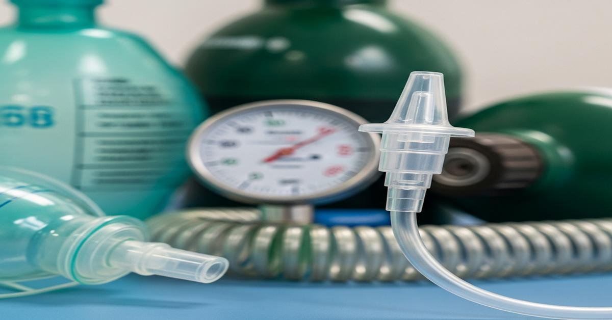
Oxygen safety and cylinder math
- Safety: Oxygen accelerates fires. Keep away from open flames and oil/grease. Secure cylinders upright. Crack the valve before attaching regulators. Green cylinders in the U.S. are oxygen.
- Safe residual pressure: Use 200 psig as your minimum in transport.
- Cylinder factors: E = 0.28; H/K = 3.14.
- Duration formula (minutes): (psig − 200) × cylinder factor ÷ flow (L/min).
Example: E cylinder at 1800 psig running 10 L/min via NRB. (1800 − 200) × 0.28 ÷ 10 = 1600 × 0.28 ÷ 10 = 448 ÷ 10 = ~44.8 minutes. Plan transport accordingly.
Home oxygen qualification (common exam points): Resting PaO2 ≤ 55 mmHg or SpO2 ≤ 88% on room air. Or PaO2 56–59 mmHg/SpO2 89% with pulmonary hypertension, cor pulmonale, or hematocrit ≥ 56%.
Airway assessment and basic maneuvers
- Assess quickly: Look for chest rise, listen for air movement and stridor, feel for airflow. Watch mental status and accessory muscle use.
- Open the airway: Head-tilt/chin-lift if no trauma. Jaw thrust if you suspect C-spine injury.
- Positioning: Sniffing position aligns the airway and eases BVM ventilation.
- Oxygen first: While you prepare devices, apply oxygen and call for help early if you anticipate difficult ventilation.
Oropharyngeal and nasopharyngeal airways
- OPA: For unconscious patients without a gag. Size from mouth corner to angle of jaw. Insert upside down then rotate, or use a tongue depressor. Why: Prevents tongue obstruction.
- NPA: For patients with an intact gag who need airway support. Size from nose to earlobe. Lubricate and insert bevel toward the septum. Avoid with facial or basilar skull fractures. Why: Better tolerance than OPA in semi-conscious patients.
Suctioning essentials
- Indications: Visible secretions, coarse breath sounds, increased work of breathing, decreased SpO2, or ventilator alarms indicating high resistance.
- Catheter size: French size ≈ (ETT ID mm × 3) ÷ 2.
- Pressures: Adults −100 to −120 mmHg; children −80 to −100; infants −60 to −80.
- Steps: Preoxygenate with 100% oxygen. Insert catheter without suction. Apply suction while withdrawing, rotating catheter. Limit to < 15 seconds (shorter for peds). Reoxygenate and reassess.
- Closed suction: Use for ventilated patients to maintain PEEP and reduce infection risk.
Bag-valve-mask ventilation (BVM)
- Seal: Two-person technique is best: one holds the mask with a two-hand “EC” grip while the other squeezes the bag.
- Oxygen supply: Use a reservoir and 15 L/min to deliver ~90–100% FiO2. Without a reservoir, FiO2 falls significantly.
- Ventilation rate: Adult 10–12 breaths/min if assisting; during CPR with advanced airway, 1 breath every 6 seconds. Give just enough volume to see chest rise. Why: Hyperventilation increases intrathoracic pressure, lowers venous return, and worsens outcomes.
- PEEP valve: Add 5–10 cmH2O if oxygenation is poor. Why: PEEP recruits alveoli and improves V/Q matching.
- Confirmation: Watch chest rise, auscultate, and monitor ETCO2 if available.
Endotracheal intubation essentials
- Indications: Inability to protect airway, apnea or severe hypoventilation, refractory hypoxemia, severe trauma, or anticipated clinical course needing airway control.
- Preparation: Preoxygenate with 100% for 3–5 minutes or 8 vital capacity breaths. Why: Extends safe apnea time.
- Tube sizes (oral): Adult female 7.0–7.5 mm; adult male 8.0–8.5 mm. Pediatrics (cuffed): size ≈ age/4 + 3.5. Depth (oral) ≈ 21–23 cm at teeth (21 women, 23 men). Peds depth ≈ 3 × ETT size (cm).
- Blades: Miller for infants/epiglottis lift; Macintosh for adults/vallecula.
- Confirm placement: Continuous waveform capnography is best. Also check bilateral chest rise and breath sounds, absent epigastric sounds. Secure depth and obtain a chest X-ray (tip 3–5 cm above carina).
- Cuff pressure: Maintain 20–30 cmH2O. Why: Prevents tracheal injury and microaspiration.
- Complications to watch: Right mainstem intubation (unilateral breath sounds), esophageal intubation, hypotension after induction, and aspiration.
Supraglottic devices: fast rescue options
- LMA/i-gel: Use when BVM is inadequate and intubation is delayed or difficult. Not definitive for aspiration risk but buys time and improves oxygenation.
Noninvasive ventilation and HFNC
- CPAP: Improves oxygenation by recruiting alveoli. Good for cardiogenic pulmonary edema and hypoxemic respiratory failure without hypercapnia.
- BiPAP (NIV): Adds inspiratory pressure support to reduce PaCO2 and work of breathing. Good for COPD exacerbation with hypercapnic acidosis.
- HFNC: For hypoxemia with high inspiratory demand or intolerance of masks. Improves comfort, provides high FiO2, and small PEEP.
- When to intubate: Worsening mental status, inability to protect airway, rising PaCO2 with acidosis, or persistent hypoxemia despite optimized support.
Mini-scenarios you must master
- 1) COPD patient, SpO2 86%, mild distress: Start Venturi 28% or NC 2 L/min. Target 88–92%. Reassess ABG if mental status worsens. Why: Avoid sudden high FiO2 that can increase PaCO2.
- 2) Trauma patient, SpO2 78%, tachypneic: Apply NRB at 15 L/min immediately. Prepare BVM and suction. If no improvement or mental status declines, assist with BVM and consider intubation. Why: Severe hypoxemia needs rapid high FiO2 and backup airway plan.
- 3) House fire victim, headache, SpO2 99% on room air: Give 100% oxygen via NRB or BVM. Send co-oximetry. Why: SpO2 is unreliable with CO; treat presumptively.
- 4) Post-op patient with atelectasis, SpO2 90% on 2 L NC: Coach IS, mobilize, and consider CPAP if oxygen need rises. Why: Oxygen does not re-expand alveoli; PEEP and deep breaths do.
- 5) Intubated patient with thick secretions and desaturation: Preoxygenate, perform closed suction with correct pressure and catheter size. Reassess SpO2 and breath sounds. Why: Retained secretions worsen V/Q mismatch and increase airway resistance.
High-yield pitfalls to avoid
- Running a simple mask below 6 L/min (risk of CO2 rebreathing).
- Ignoring dry gas at higher flows (expect nosebleeds and mucus plugging).
- Believing SpO2 in CO poisoning (it lies; use 100% oxygen and co-oximetry).
- Overventilating with BVM (causes gastric inflation and hemodynamic compromise).
- Not checking cuff pressure (tracheal damage or leaks result).
Quick daily practice checklist
- Set an SpO2 target based on the patient’s condition and stick to it.
- Choose the simplest device that meets the goal; escalate fast if it fails.
- Humidify when indicated; reassess comfort and secretions.
- For any deterioration, think airway first, then breathing, then circulation.
- Use waveform capnography whenever an advanced airway is present.
- Document device, flow/FiO2, patient response, and your plan to titrate.
- For transports, calculate cylinder duration before you roll.
Master these basics and you will answer most CRT exam airway and oxygen questions correctly. More importantly, you will make safer choices at the bedside. Start simple, reassess often, and always know why you’re choosing a device or maneuver. That is the mark of a strong therapist.

