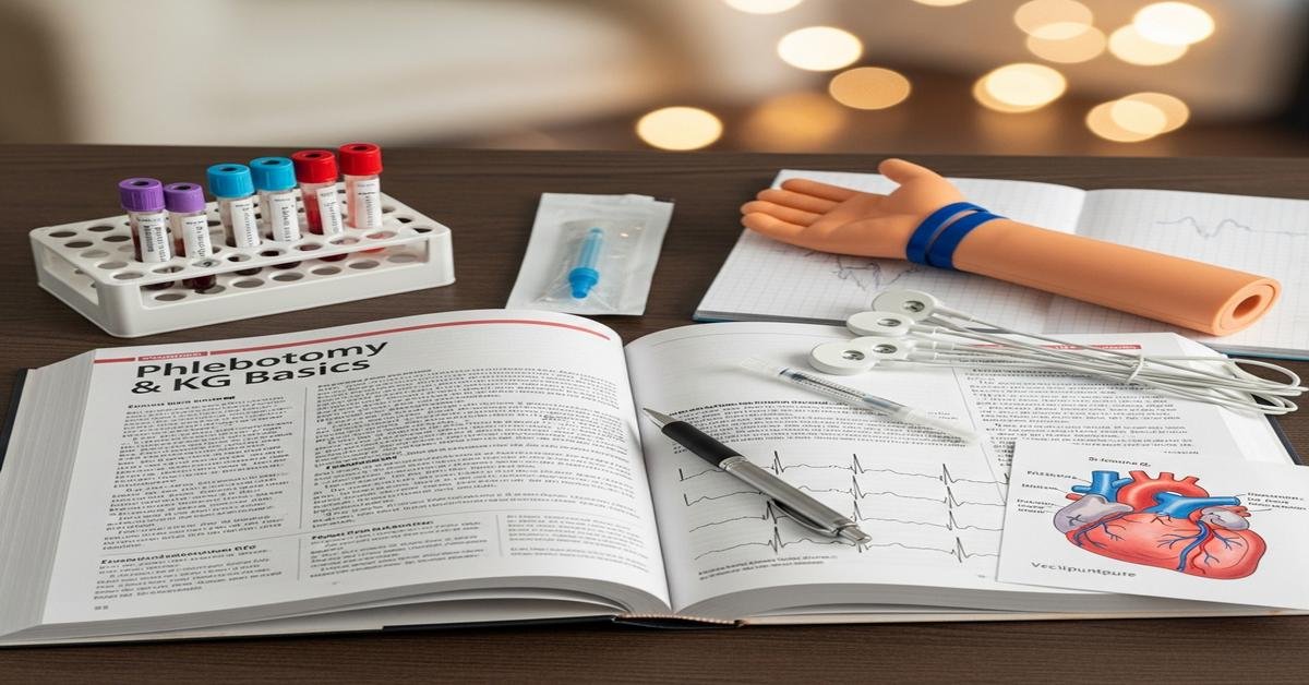
The CCMA exam expects you to be safe, precise, and consistent in two high-impact areas: phlebotomy and EKG. This guide focuses on the tasks you’ll do most often, the steps you cannot miss, and the reasons behind them. You’ll learn what the test is looking for, how to avoid common mistakes, and how to think through tricky questions the way a clinical medical assistant should.
What the CCMA Exam Expects on Phlebotomy and EKG
The CCMA doesn’t reward memorization alone. It checks that you can follow standards, protect patients, and handle variations. You’ll see scenario questions that force you to choose the safest next step, the correct order, or the best troubleshooting move.
- Phlebotomy: patient identification, order of draw, special collections, specimen handling, and complication management. The exam tests process and safety because wrong steps can invalidate tests or harm patients.
- EKG: standardization, lead placement, artifact troubleshooting, and basic rhythm recognition. Proper placement and clean tracings prevent misdiagnosis and repeat tests.
Phlebotomy Fundamentals That Score Points
1) Identify the patient every time
- Use two identifiers: full name and date of birth (or medical record number). Ask the patient to state them, then match to the order and wristband. This prevents wrong-patient errors.
- Confirm test requirements: fasting, medication timing, posture. This matters because food, position, and drugs can alter lab values.
- Explain the procedure and get consent. Anxiety and surprises increase risk of fainting and poor cooperation.
2) Choose the right site and equipment
- Preferred veins: median cubital first, cephalic second, basilic last (near nerves and artery). This reduces risk of injury.
- Avoid: the side of a mastectomy, areas with edema, hematomas, fistulas, or burns. These sites raise infection risk and give unreliable results.
- Needles: 21–22 gauge for routine draws; 23 gauge butterfly for small or fragile veins (but higher hemolysis risk). Choose the largest comfortable gauge for better flow.
3) Venipuncture steps you must do in order
- Wash hands. Apply gloves. Place the tourniquet 3–4 inches above the site for no more than 1 minute. Longer causes hemoconcentration and false high lab values.
- Clean with 70% alcohol in circles, let it air-dry. Alcohol must dry to prevent hemolysis and stinging.
- Anchor the vein. Insert the needle bevel up at 15–30 degrees (10–15 for a butterfly). A shallow, steady angle prevents transfixing the vein.
- Once blood flows, release the tourniquet before removing the needle. This reduces bruising and prevents blood from forcing out of the puncture site.
- Fill tubes in the correct order, invert as required, label at bedside, and check the patient before they leave.
4) Order of draw and why it matters
Order prevents additive carryover that can alter results. Follow this sequence:
- 1. Blood cultures (bottles or yellow SPS) – invert 8–10 times
- 2. Light blue (sodium citrate, coagulation) – 3–4 inversions; must be filled completely for correct 9:1 ratio
- 3. Red/Gold/Tiger (serum: no additive or clot activator/gel) – red glass: no inversion; SST: ~5 inversions
- 4. Green (heparin) – 8–10 inversions
- 5. Lavender/Pink (EDTA) – 8–10 inversions
- 6. Gray (fluoride/oxalate) – 8–10 inversions
Why: Citrate tubes must be collected before serum to avoid clot activator contamination. EDTA last because it chelates calcium and can ruin other tests if carried over.
5) Special collections and timing
- Blood cultures: Clean skin with chlorhexidine or iodine and let it dry. Clean bottle tops too. If using a butterfly, fill the aerobic bottle first (air in tubing would enter anaerobic bottle otherwise). This reduces contamination and false positives.
- Glucose tolerance test (GTT): Verify fasting. Draw baseline, then time starts when the patient finishes the drink. Draw exactly at ordered intervals. Small timing errors give wrong interpretations.
- Therapeutic drug levels: Confirm trough (right before dose) or peak (after dose) time. Incorrect timing invalidates results.
- Temperature/light sensitive: Ammonia and lactic acid on ice; bilirubin and folate protected from light; cold agglutinin kept warm. This preserves analyte stability.
- Drawing from an IV arm: Avoid if possible. If necessary, draw below the IV after pausing infusion for at least 2 minutes and discard 5–10 mL as waste to avoid dilution.
6) Capillary (dermal) puncture
- Sites: adult/child finger (lateral side of middle or ring finger); infant heel (medial or lateral plantar surface only). Avoid the arch and toes to protect bone and nerves.
- Depth: heel ≤2.0 mm; finger typically ≤2.0–2.5 mm depending on device. Deeper sticks risk bone injury.
- Order of draw (capillary): blood gases, EDTA, other additives, then serum. EDTA early prevents clotting and platelet clumping.
- Warm the site, clean, let dry, wipe away the first drop (tissue fluid), then collect. Gentle pressure only; squeezing causes hemolysis and tissue contamination.
7) Infection control and sharps safety
- Use standard precautions. PPE depends on exposure risk. This protects both you and the patient.
- Never recap needles. Activate safety features and dispose in a puncture-resistant sharps container immediately.
- For a needlestick or exposure: wash the area, flush mucous membranes with water, report immediately, and follow post-exposure protocol. Early action reduces infection risk.
8) Labeling and specimen handling
- Label at the bedside with two identifiers, date/time, and your initials. Labeling later causes misidentification risk.
- Let serum tubes clot upright for about 30 minutes before centrifugation unless using rapid serum tubes. Spinning early leads to fibrin clots and repeat testing.
- Transport within required time and temperature. Delays can change analyte levels.
9) Avoiding hemolysis and other errors
- Use the right needle gauge. Too small causes cell rupture.
- Let alcohol dry. Do not shake tubes; invert gently. Rough handling destroys cells and skews potassium, LDH, and more.
- Limit tourniquet time and avoid fist pumping; both concentrate blood and alter electrolytes.
- Watch for syncope. If the patient looks pale or says they feel faint, stop, remove the tourniquet and needle, and protect the patient from falls.
EKG Basics You Must Master
1) Standardization and paper settings
- Speed: 25 mm/second. Each small box = 0.04 s; large box = 0.20 s. You need this to measure intervals.
- Gain: 10 mm = 1 mV. The calibration box should be 10 mm tall. Wrong gain makes waves look abnormally large or small.
2) Lead placement: limb and chest
- Limb leads: RA (right arm) white, LA (left arm) black, RL (right leg) green, LL (left leg) red. Keep them on fleshy areas, not bone.
- Chest leads:
- V1: 4th intercostal space (ICS), right sternal border
- V2: 4th ICS, left sternal border
- V3: midway between V2 and V4
- V4: 5th ICS, midclavicular line
- V5: level with V4, anterior axillary line
- V6: level with V4, midaxillary line
- For women, place V3–V6 under the breast, not on it, to keep electrodes flat on skin. For obesity or barrel chest, landmark carefully by palpating ribs and sternum.
- If limb tremors or amputations prevent normal placement, put limb electrodes on the torso in equivalent positions and document it. Consistency keeps axis accurate.
3) Clean tracings: artifact troubleshooting
- Wandering baseline (up and down drift): check loose electrodes, skin oils, hair, and breathing movement. Clean and dry skin, shave as needed, and secure electrodes.
- Somatic tremor (shaky baseline): warm the patient, support arms/legs, ask for stillness. Tremors mimic atrial fibrillation if severe.
- AC interference (uniform 60-cycle spikes): move cords away from power sources, ensure proper grounding, and avoid crossing lead wires.
- Limb lead reversal: negative P and QRS in lead I often signals RA/LA reversal. Fix placement and repeat; wrong reversal can mimic axis deviation.
4) Quick rhythm analysis you’ll use on the exam
- Rate: 300-150-100-75-60-50 method between large boxes, or count QRS in 6 seconds and multiply by 10. This gives heart rate fast.
- Rhythm: regular or irregular? Measure R-R intervals.
- P waves: present and upright in lead II? One P for each QRS? This tells you if rhythm is sinus and if AV conduction is intact.
- Intervals:
- PR: 0.12–0.20 s (3–5 small boxes)
- QRS: ≤0.10 s is normal (up to 0.12 acceptable); wide suggests bundle branch block or ventricular rhythm
- QT: varies with rate; prolonged QT increases risk of dangerous arrhythmias
5) Common rhythms and what to look for
- Normal sinus rhythm: rate 60–100, regular, P before each QRS, normal intervals.
- Sinus bradycardia: rate <60; common in athletes or during sleep; assess for symptoms.
- Sinus tachycardia: rate >100; look for causes like pain, fever, dehydration.
- Atrial fibrillation: no distinct P waves, irregularly irregular rhythm. Irregular ventricular response stands out.
- Supraventricular tachycardia (SVT): narrow QRS, very regular, often no visible P waves. Vagal maneuvers may be ordered clinically.
- Ventricular tachycardia: wide QRS, fast, regular. This is a medical emergency if sustained.
- First-degree AV block: PR >0.20 s, constant. Often benign but notable.
- Second-degree AV block Type I (Wenckebach): PR lengthens, then a beat drops.
- Second-degree AV block Type II: constant PR with dropped beats. More serious; risk of progression.
- Third-degree block: atria and ventricles beat independently; no relation between P and QRS. Often slow and unstable.
- ST changes: elevation suggests injury/MI; depression suggests ischemia. Lead location matters: V1–V4 anterior, V5–V6 lateral, II/III/aVF inferior.
6) Holter monitors and stress tests
- Holter: place electrodes on clean, dry skin; secure wires well. Teach the patient to keep a diary of symptoms and activities, avoid showering/bathing, and not to remove electrodes. Good prep reduces artifact and repeat tests.
- Stress tests (support role): verify consent, baseline vitals, correct lead placement, and patient understanding of stopping criteria (chest pain, dizziness, severe fatigue). Early reporting of symptoms prevents harm.
Test Strategy: How to Choose the Right Answer
- Safety first: If a choice reduces harm—verify identity, stop a draw when a patient feels faint, remove a contaminated needle safely—it is usually correct.
- Follow standards: Order of draw, tourniquet time <1 minute, inversion counts, and 12-lead landmarks are test favorites.
- Sequence matters: Clean, dry, insert, release tourniquet, remove needle, then apply pressure, label at bedside. Put steps in the safest order.
- Answer what is asked: If the question says “best next step,” do not jump three steps ahead.
- Eliminate extremes: Answers that ignore precautions (“continue drawing although the patient is dizzy”) are wrong.
- Watch for distractors: For blood cultures, skin prep and drying time are more critical than needle gauge.
Quick Checklists to Review Before Exam Day
Phlebotomy essentials
- Two identifiers every time; confirm test requirements (fasting, timing).
- Preferred veins: median cubital → cephalic → basilic last.
- Avoid: side of mastectomy, edema, hematoma, burns, fistula; avoid drawing above an active IV.
- Tourniquet on <1 minute; clean and let dry; bevel up, 15–30 degrees.
- Order of draw: cultures/SPS → light blue → red/gold → green → lavender/pink → gray.
- Inversions: citrate 3–4; SST ~5; heparin/EDTA/gray 8–10; SPS 8–10.
- Release tourniquet before needle removal; immediate pressure after removal.
- Capillary: finger (adult/child), heel (infant), depth limits; wipe first drop; capillary order: gases → EDTA → others → serum.
- Special handling: ammonia/lactate on ice; bilirubin/folate protect from light; cold agglutinin warm; GTT timing exact; trough/peak timing verified.
- Label at bedside with two identifiers; allow serum to clot before spin; transport on time and at the right temperature.
- Prevent hemolysis: correct gauge, dry alcohol, gentle inversions, no fist pumping, minimize tourniquet time.
- Sharps: never recap; activate safety; report exposures immediately.
EKG essentials
- Standard: 25 mm/s speed; 10 mm/mV gain. Verify calibration mark.
- Limb electrodes: RA white, LA black, RL green, LL red.
- Chest leads: V1 4th ICS RSB; V2 4th ICS LSB; V3 between V2–V4; V4 5th ICS MCL; V5 AAL level with V4; V6 MAL level with V4.
- Skin prep: clean, dry, shave if needed; press electrodes firmly to reduce artifact.
- Artifact fixes: tighten electrodes (wandering), warm and support limbs (tremor), separate cords from power (AC interference).
- Limb reversal clue: negative complexes in lead I. Recheck and repeat.
- Rate methods: 300-150-100-75-60-50 boxes or 6-second strip ×10.
- Intervals: PR 0.12–0.20 s; QRS ≤0.10–0.12 s; watch QT.
- Common rhythms: sinus, AFib, SVT, VT, AV blocks (Types I, II, third-degree), basic ST changes.
- Holter: secure leads, diary instructions, no bathing, avoid magnets; stress test: consent, correct placement, stop for symptoms.
Why this approach works: The CCMA exam mirrors real outpatient practice. It rewards safe technique and consistent processes that reduce redraws, contamination, and misdiagnosis. If you can explain why each step matters, you’ll choose the right answer even when questions are worded in new ways.
Final practice tip: Teach each step out loud as if training a new hire. If you can justify the order, the tool, and the placement—and fix problems when something looks wrong—you’re ready for test day and for the clinic.
