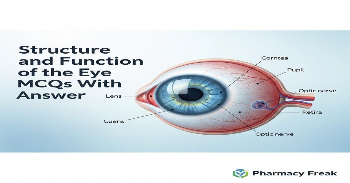
Structure and function of the eye MCQs With Answer
The structure and function of the eye are fundamental topics for B. Pharm students, linking ocular anatomy, physiology, and pharmacology. This introduction covers the cornea, lens, aqueous and vitreous humors, retina, photoreceptors (rods and cones), macula, optic nerve, and visual pathways, as well as processes like refraction, accommodation, pupillary reflexes, and intraocular pressure regulation. Understanding ocular barriers, drug entry routes, and common therapeutic targets (e.g., muscarinic, beta, prostaglandin, carbonic anhydrase) is essential for rational drug therapy in glaucoma, infections, and inflammatory eye diseases. Now let’s test your knowledge with 30 MCQs on this topic.
Q1. What is the primary refractive surface of the eye that contributes most to focusing light on the retina?
- Lens
- Cornea
- Vitreous humor
- Aqueous humor
Correct Answer: Cornea
Q2. Which structure changes curvature during accommodation to focus near objects?
- Cornea
- Ciliary muscle and lens
- Sclera
- Choroid
Correct Answer: Ciliary muscle and lens
Q3. Which layer of the retina contains the photoreceptor cells (rods and cones)?
- Ganglion cell layer
- Inner nuclear layer
- Outer nuclear layer
- Nerve fiber layer
Correct Answer: Outer nuclear layer
Q4. Rod photoreceptors are primarily responsible for which function?
- Color vision in bright light
- High-acuity central vision
- Low-light (scotopic) vision
- Stereoscopic depth perception
Correct Answer: Low-light (scotopic) vision
Q5. Which pigment is the photopigment in rods responsible for light absorption?
- Photopsin
- Melanopsin
- Rhodopsin
- Chloropsin
Correct Answer: Rhodopsin
Q6. The fovea centralis is specialized for:
- Peripheral motion detection
- High-acuity color vision
- Production of aqueous humor
- Drainage of vitreous humor
Correct Answer: High-acuity color vision
Q7. Which structure is the blind spot due to absence of photoreceptors?
- Macula lutea
- Optic disc
- Fovea
- Ciliary body
Correct Answer: Optic disc
Q8. Aqueous humor is produced mainly by which ocular structure?
- Corneal endothelium
- Ciliary epithelium
- Retinal pigment epithelium
- Lacrimal gland
Correct Answer: Ciliary epithelium
Q9. The primary route for aqueous humor drainage is through:
- Uveoscleral outflow only
- Trabecular meshwork into Schlemm’s canal
- Posterior chamber into vitreous
- Direct absorption by conjunctiva
Correct Answer: Trabecular meshwork into Schlemm’s canal
Q10. Normal intraocular pressure (IOP) range is approximately:
- 0–5 mm Hg
- 10–21 mm Hg
- 25–40 mm Hg
- 50–60 mm Hg
Correct Answer: 10–21 mm Hg
Q11. Timolol lowers intraocular pressure primarily by which mechanism?
- Increasing uveoscleral outflow
- Decreasing aqueous humor production via beta-blockade
- Inhibiting carbonic anhydrase
- Inducing miosis
Correct Answer: Decreasing aqueous humor production via beta-blockade
Q12. Pilocarpine produces miosis by acting as a:
- Beta-2 agonist
- Muscarinic receptor agonist
- Alpha-1 antagonist
- Carbonic anhydrase inhibitor
Correct Answer: Muscarinic receptor agonist
Q13. Latanoprost reduces IOP by which primary action?
- Decreasing aqueous production
- Blocking muscarinic receptors
- Increasing uveoscleral outflow via prostaglandin analog effect
- Constriction of episcleral veins
Correct Answer: Increasing uveoscleral outflow via prostaglandin analog effect
Q14. Carbonic anhydrase inhibitors (e.g., acetazolamide) decrease IOP by:
- Increasing trabecular outflow
- Reducing aqueous humor formation through bicarbonate reduction
- Contracting the ciliary muscle
- Blocking adrenergic receptors
Correct Answer: Reducing aqueous humor formation through bicarbonate reduction
Q15. Which retinal layer contains ganglion cell bodies whose axons form the optic nerve?
- Outer plexiform layer
- Inner nuclear layer
- Ganglion cell layer
- Photoreceptor layer
Correct Answer: Ganglion cell layer
Q16. In the pupillary light reflex, the afferent limb is mediated by:
- Oculomotor nerve (III)
- Optic nerve (II)
- Trochlear nerve (IV)
- Facial nerve (VII)
Correct Answer: Optic nerve (II)
Q17. The efferent limb of the pupillary light reflex that causes pupillary constriction travels in:
- Optic nerve
- Oculomotor nerve parasympathetic fibers to sphincter pupillae
- Trigeminal nerve
- Sensory fibers of the facial nerve
Correct Answer: Oculomotor nerve parasympathetic fibers to sphincter pupillae
Q18. Which statement about the blood supply of the retina is true?
- The inner retina is supplied mainly by the choroidal circulation
- The outer retina (photoreceptors) is supplied by the choroid
- The optic disc receives no blood supply
- The retina has no autoregulation
Correct Answer: The outer retina (photoreceptors) is supplied by the choroid
Q19. Retinal detachment most commonly separates which two layers?
- Retinal pigment epithelium and choroid
- Neural retina and retinal pigment epithelium
- Inner limiting membrane and vitreous
- Ganglion cell layer and inner nuclear layer
Correct Answer: Neural retina and retinal pigment epithelium
Q20. Which drug class is contraindicated when mydriasis is undesirable due to risk of precipitating acute angle-closure glaucoma?
- Topical beta-blockers
- Topical prostaglandins
- Systemic anticholinergics and topical mydriatics (e.g., tropicamide)
- Carbonic anhydrase inhibitors
Correct Answer: Systemic anticholinergics and topical mydriatics (e.g., tropicamide)
Q21. Which layer of the eye contains blood vessels that nourish the outer retina and absorbs stray light?
- Sclera
- Choroid
- Corneal stroma
- Conjunctiva
Correct Answer: Choroid
Q22. The aqueous humor flows from the posterior chamber to the anterior chamber through:
- Trabecular meshwork
- Pupil (pupillary aperture)
- Canal of Schlemm
- Vitreous cavity
Correct Answer: Pupil (pupillary aperture)
Q23. Which photoreceptor type has a faster response recovery and is responsible for color discrimination?
- Rods
- Cones
- Bipolar cells
- Horizontal cells
Correct Answer: Cones
Q24. Damage to the optic chiasm typically produces which visual field defect?
- Monocular blindness
- Bitemporal hemianopia
- Quadrantanopia
- Homonymous hemianopia
Correct Answer: Bitemporal hemianopia
Q25. Which ocular barrier most limits systemic drug penetration into the posterior segment (retina) when using topical eye drops?
- Blood-aqueous barrier
- Blood-retinal barrier
- Conjunctival epithelium only
- Lacrimal film
Correct Answer: Blood-retinal barrier
Q26. Which neurotransmitter predominates in the parasympathetic innervation of the eye?
- Norepinephrine
- Dopamine
- Acetylcholine
- Glutamate
Correct Answer: Acetylcholine
Q27. Which optical condition results from an elongated eyeball causing light to focus in front of the retina?
- Hyperopia (farsightedness)
- Emmetropia
- Myopia (nearsightedness)
- Presbyopia
Correct Answer: Myopia (nearsightedness)
Q28. Presbyopia is primarily due to age-related changes in which ocular component?
- Corneal curvature
- Lens elasticity and reduced accommodation
- Retinal photoreceptor loss
- Ciliary nerve degeneration
Correct Answer: Lens elasticity and reduced accommodation
Q29. Which enzyme cascade is directly involved in phototransduction in rods when rhodopsin is activated?
- Adenylyl cyclase → cAMP
- Guanylate cyclase → cGMP increase
- Transducin activation → phosphodiesterase → cGMP hydrolysis
- Protein kinase A activation only
Correct Answer: Transducin activation → phosphodiesterase → cGMP hydrolysis
Q30. A lesion in the right optic tract produces which visual field defect?
- Bitemporal hemianopia
- Left homonymous hemianopia
- Right monocular blindness
- Central scotoma
Correct Answer: Left homonymous hemianopia


